All Posts in Category: Procedures

SPLENECTOMY

What do you need to know about your laparoscopic splenectomy?

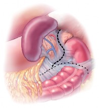
Your doctor(s) have indicated that you require surgical removal of your spleen. The following information can help explain what you can expect from a laparoscopic splenectomy. A laparoscopic or minimally invasive approach involves specialized video equipment and instruments that allow a surgeon to remove the spleen through several tiny incisions, versus a traditional large midline incision for an open approach. The benefits of minimally invasive surgery include a shorter hospital stay, a faster return to normal activity and less scarring.

The most common conditions that warrant splenectomy in an adult are:

Hereditary spherocytosis – a blood disorder characterized by numerous abnormally (sphere) shaped red blood cells.
Idiopathic thrombocytopenia purpura (ITP) – a blood disorder that involves bruising and spontaneous bleeding due to an acute shortage of platelets.
Staging for Hodgkin’s Lymphoma (cancer originating in the lymph node) – physicians will sometimes remove the spleen to determine the extent of involvement and plan of treatment.
In preparation for surgery, routine preoperative tests such as an ECG and a chest x-ray may or may not be needed. You will have already completed an extensive series of blood tests and a sonar or CT scan to establish your diagnosis. Standard protocol for spleen removal also involves vaccination against certain infections.
You will be asked to refrain from eating 8 hours before surgery. Your doctor will evaluate any medications you are taking prior to surgery and determine if any of these medications need to be discontinued.
The laparoscopic splenectomy is performed with general anaesthesia.
As with any other operation involving the abdomen, injury to abdominal organs and tissues is a potential risk. Other complications include bleeding and infection. Pre-operative tests and blood type matching or patient donation will prepare for a blood transfusion if necessary. If during the
course of the operation the surgeon needs to make a traditional incision to safely complete the operation, this should not be seen as a failure but as a wise decision to prevent dangerous complications.
Once you are asleep, the operating room team will work together to perform your operation. When your operation is complete, you will be awakened from anaesthesia in the operating room but you may not remember this. For your comfort and safety, you will be attended to in the specialised intensive care unit or High Care. After a few hours, the nurses will help you out of bed and give you something to drink. It is common to feel groggy and nauseated soon after surgery and medication is available to help with these discomforts. Although a minimally invasive approach can significantly shorten the length of your stay in the hospital, your physician will determine when you can be released based on a number of issues specific to your condition.

POST-OPERATIVE INSTRUCTIONS FOLLOWING LAPAROSCOPIC SPLENECTOMY

ACTIVITES

A laparoscopic splenectomy causes less damage to the muscles and other tissues than a standard splenectomy incision. For this reason, there are fewer restrictions on your physical activity than might be expected.
Walking: Walking is permitted and encouraged beginning within hours of your operation. Start with short walks and gradually increase the distance and length of time that you walk.
Climbing: Climbing stairs is permitted. Initially, some assistance may be necessary.
Lifting: Lifting is permitted without restrictions.
Showers: Showers are permitted two days after surgery. Be careful to clean your incision (plaster and all), with a mild soap. Rinse well and pat dry.
Driving: Driving may be resumed 7 days following surgery. Care should be taken after that point if you are still taking prescription pain medications.

DIET

There are generally no dietary restrictions following surgery. Foods which cause you discomfort or do not agree with you should be avoided. Most patients find that they are able to eat many foods that they were not able to tolerate prior to surgery. Gradually introduce these items to your
diet and determine how well you tolerate them.

MEDICATIONS

Pain: Your physician will prescribe Pain medications after surgery. We recommend Tramacet for mild to moderate pain. If this does not sufficiently control your pain, take the prescribed pain medications according to the directions on the label.
Stool Softener: Stool softener or mild laxative may be necessary if you do not have a spontaneous bowel movement within 3 days of your surgery. Call the office for further instructions.

RETURN TO WORK

Most patients will be able to return to work or resume their usual level of function 7-10 days after surgery. This may need to be determined by you and your employer. Some patients have residual fatigue for a couple of weeks following general anaesthesia.
CALL THE OFFICE if you have any questions or problems. Call immediately if you notice any of the following symptoms:

  1. Persistent nausea or vomiting.
  2. Fever greater than 380C.
  3. Increased abdominal pain.
  4. Pus or increased redness around the incisions.
  5. Severe shoulder pain lasting more than 3 days.

This information is not intended to take the place of a visit with your physician. If you have further questions about preoperative symptoms or postoperative conditions, please contact your physician.

Read More

THORACOSCOPIC SYMPHATECTOMY

GENERAL INFORMATION

A sympathectomy is the removal of some of the sympathetic nerves which are responsible for control of the perspiration from the upper limb. This is done by means of an endoscope which is inserted through the chest cavity.

COMMON SIGNS AND SYMPTOMS

The disease hyperhidrosis results in excessive perspiration usually of the hands but sometimes also of the armpits

DIAGNOSIS

  • The diagnosis is made clinically
  • There are no tests to be done to make this diagnosis.
  • Sometimes your doctor may decide to do a pre-operative chest X-ray

TREATMENT

  • No creams or medication really cure or control this condition
  • Botox injections are sometimes used but these give only temporary relief and can not cure this condition
  • Your doctor has advised you to have a thoracoscopic sympathectomy.

PRE-OPERATIVE PREPARATION

  • Make sure your doctor knows what medicines you take, especially medicines which may affect blood clotting.
  • Do not eat or drink anything for 6 hours before the operation.

OPERATION

The operation will be done under general anaesthetic. Two or rarely three small incisions will be used on each side of your chest through which cannulae are inserted.

  • An endoscope is used to view the sympathetic chain of nerves and the sympathetic ganglia at the back of the chest cavity by first collapsing the lung.
  • This is a safer way of doing the operation than the previous way of doing the procedure through the neck.
  • Two of the sympathetic ganglia are then divided or destroyed to interrupt the nerve impulses to the sweat glands of the hands. If the axilla is involved, then a third ganglion can be divided too.

POST OPERATIVE CARE

  • After the operation you will be taken to a recovery room. When you are stable you will be taken to your regular hospital bed.
  • Your doctor may decide to do a post-operative chest X-ray.
  • Any pain, discomfort, or nausea will be controlled with medication.
  • You can expect to be discharged a day or two after the operation.

COMPLICATIONS

  • Serious complications are infrequent after a thoracoscopic sympathectomy. It is however important for you to remember that it is a major operation and complications may occur.
  • Air may re-accumulate in your chest cavity after the operation forming a pheumothorax which may require a chest drain
  • A bleed or an infection is always possible at the site of the small incisions.
  • It is possible for the heart or lungs to be injured during this operation. This is rare. This may necessitate an open operation of the chest.
  • A Horner’s syndrome with a drooping eyelid is possible but rare
  • The operation may not be successful. The original hyperhidrosis can recur after months or years
  • The biggest problem after this operation is that there is mostly a measure of compensatory hyperhidrosis. One does not really have control of this. It often manifests as increased perspiration from the body, the face or from the feet.
  • Systemic complications like venous thrombosis and pulmonary embolism are rare, but possible after any operation or anaesthetic.
Read More

Morning Moviprep

Moviprep – 2 dose bowel cleaning

If your appointment is in the MORNING, follow the instructions below

The following instructions are for appointments before noon only – please check your appointment time

How to take MOVIPREP

It is essential you take two doses of the medication provided according to the following instructions (rather than the manufacturer’s leaflet). The preparation of the medication and a suggested timetable for food and medication has been provided to maximise the success of the procedure and reduce the amount of the side effects experienced.

Moviprep is a two dose treatment. The pack contains four sachets: two ‘Sachet A’s and two ‘Sachet B’s.  To make one dose, mix one of the ‘Sachet A’ with one of the ‘Sachet B’ with a litre of water. Stir until the podwer is completely dissolved and the solution is clear or slightly hazy, this may take up to five minutes. You may add cordial to improve the taste (not blackcurrant).

Each dose (1 litre) of Moviprep should be drunk slowly over a period of 2 hours. There is no need to rush, a glass (250ml) every 30 minutes is sufficient. You should also drink an additional 500mls of water or clear fluids with each litre of Moviprep taken.


REMEMBER KEEP HYDRATED – DRINK ONE CUP OF WATER (250ML) EVERY HOUR

Suggested routine to prepare for your procedure

The day before your procedure
Breakfast 7-8am

Eat breakfast as normal, avoiding high fibre foods, fruits and vegetables.

You may have milk and dairy products. This will be the last solid meal until after you procedure.

9am

From this time onwards, DO NOT EAT ANY SOLID FOOD, your bowel needs to be completely empty for your procedure. You can drink clear soups, soft drinks and black tea or coffee with sugar as you wish.

Lunch

You can have clear soups such as Bovril or Oxo, Remember to continue to drink plenty of clear fluids throughout the day.

4 – 5pm

Make your first 1 litre dose of Moviprep and drink over a period of 1 to 2 hours REMEMBER TO DRINK AN ADDITIONAL 500ml OF WATER FOLLOWING THIS DOSE.

7 – 8pm

Make your second 1 litre dose of Moviprep and drink over a period of 1 to 2 hours.   REMEMBER TO DRINK AN ADDITIONAL 500ml OF WATER FOLLOWING THIS DOSE.

11pm

Once your bowel is empty and you have stopped going to the toilet, go to bed. This is normally about 2 hours after you finish the Moviprep.

The day of your procedure

DO NOT EAT / DRINK ANYTHING, from 24h00 the previous night. Leave home in time for your appointment.

POSSIBLE SIDE EFFECTS
Everybody responds differently to bowel cleansing. We advise you to remain in easy reach of a toilet as Moviprep causes multiple, often urgent bowel movements. Moviprep can start working quickly and bowel movements are normally seen for at least two hours, however the effects can last a lot longer. Moviprep may make you feel nauseated or sick, feel tired, have abdominal bloating or cramp. The lack of solid food in combination with the Moviprep can trigger nausea. We suggest you drink plenty of clear fluid to help prevent this. The frequent bowel movements can cause anal irritation. Occasionally an allergic reaction can occur, with or without a rash, if you experience this please seek medical advice.


Please check Moviprep leaflet for side effects, contra – indications, etc.

Please take blood pressure medication as usual on morning of procedure with a small amount of water.

DO NOT take diabetic medication/ insulin the day of procedure

Please contact my rooms if you have any questions/uncertainties

Read More

Afternoon Moviprep

Moviprep – 2 dose bowel cleaning

If your appointment is in the AFTERNOON, follow the instructions below

The following instructions are for appointments between 12 noon and 6pm only– please check your appointment time

How to take MOVIPREP

It is essential you take two doses of the medication provided according to the following instructions (rather than the manufacturer’s leaflet). The preparation of the medication and a suggested timetable for food and medication has been provided to maximise the success of the procedure and reduce the amount of the side effects experienced.

Moviprep is two dose treatment. The pack contains four sachets: two ‘Sachet A’s and two ‘Sachet B’s. To make one dose, mix one of the ‘Sachet A’ with one of the ‘Sachet B’ with a litre of water. Stir until the power is completely dissolved and the solution is clear or slightly hazy, this may take up to five minutes. You may add cordial to improve the taste (not blackcurrant).
Each dose (1 litre) of Moviprep should be drunk slowly over a period of 2 hours. There is no need to rush, a glass (250ml) every 30 minutes is sufficient. You should also drink an additional 500mls of water or clear    fluids with each litre of Moviprep taken.

REMEMBER KEEP HYDRATED-DRINK ONE CUP OF WATER (250ML) EVERY HOUR

Suggested routine to prepare for your procedure


The day before your procedure

Breakfast

Eat breakfast as normal.

Launch 12 noon

Eat lunch, avoiding high fibre foods, fruits and vegetables. You may have milk and dairy products. This will be the last solid meal until after your procedure.

1pm

From this time onwards, DO NOT EAT ANY SOLID FOOD, your bowel needs to be completely for your procedure. You can drink clear soups, soft drinks and black tea or coffee with sugar as you wish.

Teatime 5pm

You can have clear soups as Bovril or Oxo, Remember to continue to drink plenty of clear fluids throughout the day.

7pm

Make your first 1 litre dose of Moviprep and drink over a period of  1 to 2 hours. REMEMBER TO DRINK AN ADDITIONAL 500 ML OF WATER FOLLOWING THIS DOSE.

10pm

Once your bowel is empty and you have stopped going to the toilet, go to bed. This is normally about 2 hours after you finished the Moviprep.

The day of your procedure 6am

Remember DO NOT EAT / DRINK ANYTHING, from 08h00. Make your second 1 litre dose of Moviprep and drink over a period of 1 to 2 hours. Remember to drink an additional 500ml of water   following this dose.

Later

YOU will have watery bowel movements for up to 2 hours after   finishing your Moviprep; allow plenty of time for your bowel to empty before leaving for your appointment.

POSSIBLE SIDE EFFECTS

Everybody responds differently to bowel cleansing. We advise you to remain in easy reach of a toilet as Moviprep causes multiple, often urgent bowel movements. Moviprep can start working quickly and bowel movements are normally seen for at least two hours, however the effects can last a lot longer. Moviprep may make you feel nauseated or sick, feel tired, have abdominal bloating or cramp. The lack of  solid food in combination with the Moviprep can trigger nausea. We suggest you drink plenty of clear fluid to help prevent this. The frequent bowel movements can cause anal irritation. Occasionally an allergic reaction can occur, with or without a rash, if you experience this please seek medical advice.

Please check Moviprep leaflet for side effects, contra – indications, etc.

Please take blood pressure medication as usual on morning of procedure with a small amount of water.

DO NOT take diabetic medication/ insulin the day of procedure

Please contact my rooms if you have any questions/uncertainties

Read More

SPLENECTOMY

What do you need to know about your laparoscopic splenectomy?
Your doctor(s) have indicated that you require surgical removal of your spleen. The following information can help explain what you can expect from a laparoscopic splenectomy.
A laparoscopic or minimally invasive approach involves specialized video equipment and instruments that allow a surgeon to remove the spleen through several tiny incisions, versus a
traditional large midline incision for an open approach. The benefits of minimally invasive surgery include a shorter hospital stay, a faster return to normal activity and less scarring.
The most common conditions that warrant splenectomy in an adult are:
Hereditary spherocytosis – a blood disorder characterized by numerous abnormally (sphere) shaped red blood cells.
Idiopathic thrombocytopenia purpura (ITP) – a blood disorder that involves bruising and spontaneous bleeding due to an acute shortage of platelets.
Staging for Hodgkin’s Lymphoma (cancer originating in the lymph node) – physicians will sometimes remove the spleen to determine the extent of involvement and plan of treatment.
In preparation for surgery, routine preoperative tests such as an ECG and a chest x-ray may or may not be needed. You will have already completed an extensive series of blood tests and a
sonar or CT scan to establish your diagnosis. Standard protocol for spleen removal also involves vaccination against certain infections.
You will be asked to refrain from eating 8 hours before surgery. Your doctor will evaluate any medications you are taking prior to surgery and determine if any of these medications need to be
discontinued.
The laparoscopic splenectomy is performed with general anaesthesia.
As with any other operation involving the abdomen, injury to abdominal organs and tissues is a potential risk. Other complications include bleeding and infection. Pre-operative tests and blood
type matching or patient donation will prepare for a blood transfusion if necessary. If during the course of the operation the surgeon needs to make a traditional incision to safely complete the
operation, this should not be seen as a failure but as a wise decision to prevent dangerous complications.
Once you are asleep, the operating room team will work together to perform your operation. When your operation is complete, you will be awakened from anaesthesia in the operating room
but you may not remember this. For your comfort and safety, you will be attended to in the specialised intensive care unit or High Care. After a few hours, the nurses will help you out of
bed and give you something to drink. It is common to feel groggy and nauseated soon after surgery and medication is available to help with these discomforts. Although a minimally
invasive approach can significantly shorten the length of your stay in the hospital, your physician will determine when you can be released based on a number of issues specific to your condition.
POST-OPERATIVE INSTRUCTIONS FOLLOWING LAPAROSCOPIC SPLENECTOMY ACTIVITES
A laparoscopic splenectomy causes less damage to the muscles and other tissues than a standard splenectomy incision. For this reason, there are fewer restrictions on your physical activity than
might be expected.
Walking: Walking is permitted and encouraged beginning within hours of your operation. Start with short walks and gradually increase the distance and length of time that you walk.
Climbing: Climbing stairs is permitted. Initially, some assistance may be necessary.
Lifting: Lifting is permitted without restrictions.
Showers: Showers are permitted two days after surgery. Be careful to clean your incision (plaster and all), with a mild soap. Rinse well and pat dry.
Driving: Driving may be resumed 7 days following surgery. Care should be taken after that point if you are still taking prescription pain medications.
DIET
There are generally no dietary restrictions following surgery. Foods which cause you discomfort or do not agree with you should be avoided. Most patients find that they are able to eat many
foods that they were not able to tolerate prior to surgery. Gradually introduce these items to your diet and determine how well you tolerate them.
MEDICATIONS
Pain: Your physician will prescribe Pain medications after surgery. We recommend Tramacet for mild to moderate pain. If this does not sufficiently control your pain, take the prescribed pain
medications according to the directions on the label.
Stool Softener: Stool softener or mild laxative may be necessary if you do not have a spontaneous bowel movement within 3 days of your surgery. Call the office for further instructions.
RETURN TO WORK
Most patients will be able to return to work or resume their usual level of function 7-10 days after surgery. This may need to be determined by you and your employer. Some patients have
residual fatigue for a couple of weeks following general anaesthesia.
CALL THE OFFICE if you have any questions or problems. Call immediately if you notice any of the following symptoms:
1. Persistent nausea or vomiting.
2. Fever greater than 380C.
3. Increased abdominal pain.
4. Pus or increased redness around the incisions.
5. Severe shoulder pain lasting more than 3 days.
This information is not intended to take the place of a visit with your physician. If you have
further questions about preoperative symptoms or postoperative conditions, please contact your
physician.

Read More

MALIGNANT MELANOMA

Melanoma is a type of cancer which usually starts in the skin. It is a cancer that grows from melanocytes – the skin cells which produce the protective pigment called melanin. Since most melanocytes are found in the skin, this is the commonest site for melanoma. Many but not all melanomas grow from moles.

Understanding the structure of the skin helps to show how melanoma develops.

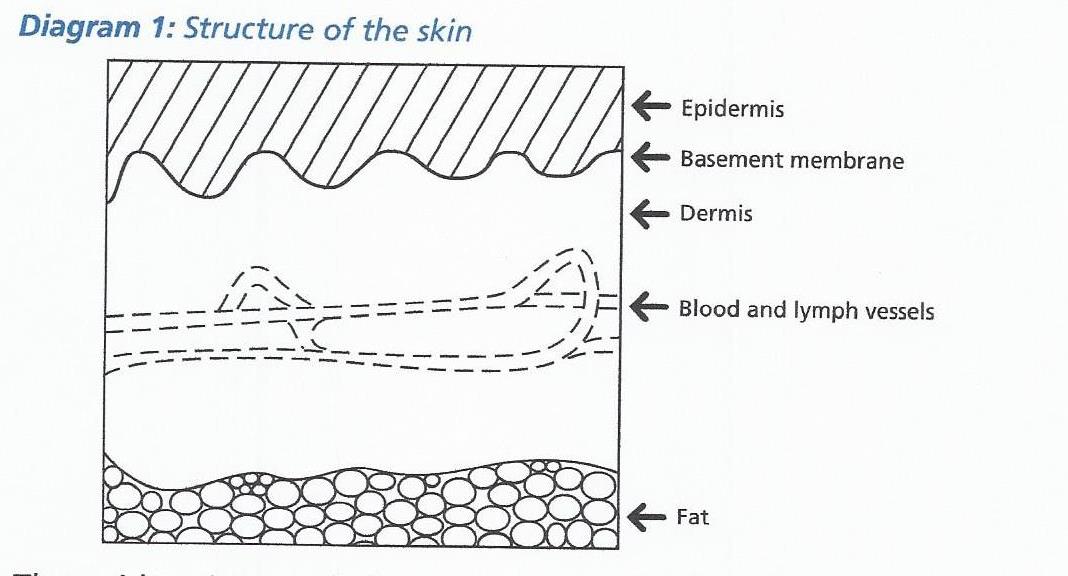
The skin consists of a thin protective outer layer called the epidermis, and a thicker inner layer, the dermis. Each layer is made of individual “building blocks” called cells, and the two layers are separated by a boundary – the basement membrane.
MALIGNANT MELANOMA - 1


The epidermis is made from two types of cells, called keratinocytes and melanocytes. The keratinocytes make up most of the epidermis, whereas the melanocytes make the pigment that causes the skin to look brown. This pigment is called melanin, and protects the skin from sunburn. The second, inner layer, the dermis, contains blood and lymphatic vessels, which together form the ‘plumbing’ system in the skin.

Melanoma some useful facts:

The skin is constantly wearing out and replacing itself by making new cells. Sometimes things go wrong and the cells replace themselves too quickly. This results in a tumour, which is simply a lump of abnormal cells. Tumours can be benign or malignant.

Benign tumours are usually quite small, they do not grow into the surrounding tissues, and they do not spread elsewhere in the body. For example a wart is a harmless growth of the keratinocytes, and a mole is a harmless growth of the melanoytes in the skin. Benign tumours are not cancer.

Malignant tumours can invade and destroy surrounding tissues and may spread or ‘metastasise’ to other parts of the body. Malignant tumours are cancer.

It is important to remember that cancer is not one disease; it is a group of many diseases. Each type of cancer differs from the others in many ways and the diagnosis, treatment and follow up varies between types of cancer.

Cause of melanoma

The precise cause of melanoma is not fully understood. However there is evidence to suggest that ultraviolet radiation from the sun may damage the skin and cause melanoma. In general the risk is greatest in those with white skin, especially those who burn easily and tan poorly. It is also greater in those with a large number of moles. Melanoma can be inherited but this is very uncommon.


Types of melanoma

Melanomas in the skin occur in two main forms:

  1. In-situ melanoma
  2. Invasive melanoma

In-situ melanoma :

This type accounts for about 10% of the melanomas. The most important point about in-situ melanoma is that it is harmless if treated correctly. The cancer cells are confined to the top layer of the skin, the epidermis. They are separated from the blood vessels and lymphatic vessels by the basement membrane, as shown in the picture. However, if untreated,  in-situ melanoma can continue to grow and break through the membrane and could then potentially spread. For this reason it is important that they are treated.

Invasive melanoma:

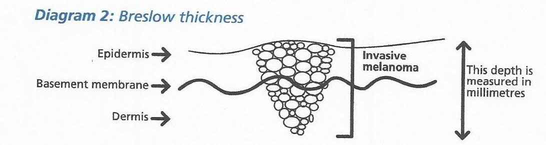
Invasive melanomas are not confined to the top layer of the skin. They grow through the basement membrane into the deeper layer, the dermis. Here, blood and lymph vessels are present which can provide a route for cancer cells to spread around the body. The further down a melanoma has grown into the skin, the greater the chance of the cancer cells getting inside a blood or lymph vessel, and being carried away from the skin to another part of the body. Invasive melanoma is therefore more serious than in-situ melanoma, because the cancer has the potential to spread.

The single most important point about your melanoma is therefore the depth to which it has invaded into the skin, the greater the depth or thickness, the greater the risk of spreading. This measurement is made very carefully on each melanoma that we remove. The measurement is made by an expert pathologist using a microscope. It is measured in millimetres and is referred to as the Breslow thickness (diagram 2).
 MALIGNANT MELANOMA - 2

It is important to realise that for most melanomas the chance of spreading is small, because most melanomas remain close to the surface.

The thinner the melanoma, the smaller the chance of it having spread before it was removed, and the greater the chance of cure. If you want to know more about this in detail please ask your Consultant. However, it is important to remember that about 80% of all patients with melanoma are cured.


TREATMENT

 Biopsy

Where possible the entire mole or lesion is removed under local anaesthetic, with a margin of normal skin so that it is completely removed. The specimen is sent to the pathology department where it is examined carefully. If it is a melanoma, its thickness, the level of penetration into the skin, and some other features are looked at. This helps us to decide the most appropriate treatment for you.

Melanoma is primarily treated with surgery. Surgical treatments can be divided into three main categories:

  1. Treatment of the primary melanoma,
  2. Treatment of the lymph glands
  3. Treatment of metastases (spread of melanoma). A detailed explanation of the new drug treatments is found further down this page.

PRIMARY MELANOMA

WHY DOES MY SMALL MELANOMA NEED SUCH A BIG EXCISION?

When melanomas grow on the surface of the skin they enlarge in diameter. This happens because the melanoma cells have the ability to move along the surface of the skin. Some melanoma cells move more quickly than others and spread further away from the main tumour. This results in a melanoma which is surrounded by individual scattered cells that are not visible. The deeper a melanoma has penetrated into the skin the wider the scatter on the surface. When treating the primary melanoma it is necessary to ensure that all the invisible scattered cells are removed otherwise the melanoma will re-grow in the same site. This is done by excising (cutting out) a safety margin around the biopsy site of the primary melanoma. This is termed a wide local excision.

The size of safety margin is determined by the depth of the original melanoma because the deeper the melanoma, the wider the scatter effect on the surface. Thin melanomas need a 0.5 cm margin while thicker melanomas require a 2 cm margin of safety. Most of the time the defect that is created with a wide local excision can be closed with a skin flap. In the lower leg, however, there is often not enough stretch in the surrounding skin to be able to do a flap so one has to resort to doing a skin graft.

  1. LYMPH GLANDS

WHY DO I NEED ALL MY SURROUNDING LYMPH GLANDS REMOVED?

Sentinel lymph node biopsy (SLNB)

When melanomas spread they often do so via the lymph vessels to the lymph glands. For instance a melanoma on the leg would be expected to spread to the lymph glands in the groin. The first lymph gland to be affected is called the sentinel lymph node. It can be identified and removed to test whether the melanoma has spread. It enables us to find out if there has been any spread to the lymph nodes at the time of diagnosis and gives us a more accurate way to stage your disease.

If there is a confirmed metastasis to a lymph node (either an enlarged gland or a positive gland found on sentinel node biopsy) then it is necessary to remove all the remaining lymph nodes from that area (called a therapeutic node dissection or block dissection). This is done because we know that if one gland is affected in an area then there is between a 20% and 40% chance that other glands are also involved. This surgery is performed under general anaesthesia, usually takes about 1-2 hours and patients are required to stay in hospital for one night. If there has been no further spread in the blood, therapeutic node dissection provides an opportunity for cure of the melanoma.

Risks and benefits

As with any surgery there is a risk of bleeding and infection, you may experience some pain and you will be left with a scar. If there is any other risks relating to your surgery the surgeon will discuss this with you before treatment.

After surgery

After your operation you will be given information about any stitches that need to be removed, any dressing changes that are needed and be informed about your follow-up appointment at the rooms. Again this time varies from person to person and may be the following day or could be 2 weeks later.

At this appointment, your wounds will be checked and the pathological results and further management will then be discussed – you may be referred for oncological opinion/management if appropriate.

  1. SYSTEMIC METASTASES

MY MELANOMA HAS SPREAD. WHAT TREATMENTS ARE AVAILABLE?

If a melanoma has managed to spread in the blood stream to a distant organ (systemic metastasis) surgery remains the best chance of a cure. Even if a cure is not achieved, the survival time with surgery is significantly longer than that obtained with a combination of treatment drugs and radiotherapy. Where possible, surgery is offered as a treatment for spread to organs. Only if surgery is not technically possible or if there are too many sites of spread or one has a high risk of developing metastases, does one consider the need for treatment drugs or radiotherapy.


RADIOTHERAPY:

In terms of radiotherapy, radiation can be used in the adjuvant setting after surgery. When a melanoma is in a clinical site that is difficult to get a wide clearance and adequate margins after surgery, then radiation can be used postoperatively if the wide local excision is narrower than the ideal. Patients sometimes have to undergo removal of lymph glands from certain anatomical sites such as the groin or under the arm and when certain criteria are met, such as many involved nodes or extracapsular spread, patients require postoperative radiation to these areas in order to reduce the risk of local recurrence. This radiation does not improve survival but rather local control. Radiation also plays an important role in palliation or relief of symptoms in patients who have advanced or metastatic disease where the cancer has spread to other sites within the body. It can be used for pain relief or to reduce pressure on structures or to reduce bleeding. Radiation is generally given on a daily basis on weekdays and takes about 15 to 20 minutes to administer and the treatment is aimed and targeted at the area of interest.

NEW DRUG TREATMENTS FOR MELANOMA

All the new treatments for metastatic melanoma are very expensive and most are not covered by medical aids. Only one of the six drugs discussed below has been released for use in South Africa ,namely Ipilimumab. The average cost of each drug is about R1 million per year. The optimal length of treatment for each of the drugs has not been established so no one knows how long they need to be taken for. Some of these drugs have a lot of side effects which forces people to stop taking them and the number of side effects often increases when one combines drugs. Otherwise, the new drugs for treating metastatic melanoma have been able to produce some spectacular results and have brought a lot of hope to many people. There is evidence that some patients may actually be cured of metastatic melanoma by these new drugs. 

Research is ongoing and is actually accelerating in the field of biological drugs, so one would predict that many new, more effective and possibly cheaper drugs may be available in the coming years.

SUMMARY OF THE NEW DRUG TREATMENTS FOR MELANOMA

There are two main groups of drugs (all discussed in detail below)

  1. Chemical drugs that block mutated enzymes in melanoma cells. The mutated enzymes send signals for the melanoma cells to divide and spread. When the enzymes are switched off, the melanoma cells die.
  • Vemurafenib
  •  Dabrafenib
  •  Trametinib
  •  Combination therapy with Dabrafenib and Trametinib


  1. Antibodies that stimulate the body’s own immune system to kill melanoma cells.
  •  CTLA-4 inhibitors – Ipilimumab
  •  PD1 inhibitors – Nivolumab
  • Pembrolizumab
  • Combination therapy with both Nivolumab and Ipilimumab (approved by the FDA in October 2015)


HOW THE IMMUNE SYSTEM WORKS

The main cells of the immune system that are responsible for killing cancer cells are called T-lymphocytes (They get their name from the fact that they are mostly found in lymph tissue. They are called T lymphocytes because they mature in the Thymus gland in the neck). T-lymphocytes cannot immediately recognise cancer cells and therefore have to first be “taught” to do so by other cells. The cells that do the teaching are called accessory or antigen presenting cells. These are very large (but not very numerous) cells that have an innate ability to recognise tumour cells as being foreign. When an accessory cell encounters a cancer cell it is able to ingest (phagocytose) this cell and then destroy it by enzymatically breaking it up into lots of little particles (called antigens). The accessory cell then takes the tumour particles and pushes them through onto the surface of its cell membrane where they become attached to proteins called MHC (major histocompatibility complex protein). The accessory cell then shows or “presents” the tumour particles (antigen) combined with the MHC protein to the T-Lymphocytes (hence the accessory cell’s other name of antigen presenting cell). In this way it shows the T-lymphocytes parts of the destroyed cancer cell and thus “teaches” the T-lymphocytes to recognise the cancer cells that need to be killed. The T-lymphocyte actually binds to the surface of the accessory cell for this process to happen. Once released from the accessory cell, the T-lymphocyte has become activated to go and hunt for cancer cells, which it kills by ingestion (phagocytosis).

An interesting thing happens when the T-lymphocyte is attached to the accessory cell. As well as activating the T-lymphocyte cell, the accessory cell also initiates a process to begin inactivation of the T lymphocyte. It does this by switching on a protein on the T-lymphocyte surface called CTLA-4 (cytotoxic T-lymphocyte-antigen 4). Initially this CTLA-4 (off switch) is not very active but over time it becomes more so until it is so active that the T-lymphocyte switches off. The body does this as a control mechanism so that there is a way of regulating the active T-lymphocyte cells. Unfortunately, the switch-off mechanism is often premature and occurs before all the cancer cells have been killed. Once this switch-off mechanism had been identified, intense research resulted in the production of a drug which allowed the switching off mechanism to be blocked. An antibody was produced which binds to the CTLA-4 (off switch) molecule prior to T-lymphocyte binding to the accessory cell. This thus prevents the CTLA-4 (off switch) from being activated by the accessory cell. T-lymphocytes thus become active for much greater periods of time and can kill a far larger number of cancer cells. The name given to the antibody blocker is Ipilimumab.

Further research has found that active T-lymphocytes have yet another surface molecule that allows them to be switched off. The molecule is called PD1 (programmed death receptor 1). Healthy human cells need to be able to protect themselves from active T-lymphocytes so they all have a protein on their cell surfaces called PD-L (programmed cell death ligand). When an active T-lymphocyte comes along and tries to attack a healthy cell, the healthy cell binds its PD-L molecule with the active T-lymphocyte PD1 molecule and the T-lymphocyte gets switched off. The unfortunate thing is that many cancer cells also carry the PD-L molecule. They can therefore also inactivate the T-lymphocytes and protect themselves from destruction. Science has again stepped in to produce an antibody which, when given intravenously, attaches to the PD1 receptor on the surface of the T-lymphocyte. The function of the antibody is to get in the way and make it impossible for the PD-L molecule on cancer cells to attach to the PD1 receptor on the T-lymphocyte. The active T-lymphocyte therefore cannot be switched off. There are two antibodies that have been produced to block PD1 receptors known as Nivolumab and Pembrolizumab.

  1. Ipilimumab

– This is a CTLA-4 blocker (blocks the off switch)
– Approved by the FDA in March 2011 for the treatment of metastatic melanoma.
– Sold under the trade name of Yervoy
– One of the big problems with Ipilimumab is that only about 10% of patients have a response to the drug.
– Another problem with Ipilimumab is that about 27% of patients experience severe side effects.
– The good news, however, is that some people appear to have been cured by Ipilimumab with a survival rate of 9% at ten years. – So far, no other treatment for metastatic melanoma can boast such good results.
– Ipilimumab is given as an intravenous infusion initially once every three weeks for four times and can be repeated if there is progression of disease more than three months after an initial response.

  1. Pembrolizumab (Pembro)

– This was the first of the PD1 inhibitors to be approved by the FDA for treating metastatic melanoma. Approval for use given by the FDA in September 2014.
– It is sold under the trade name Keytruda.
– The average number of patients who respond to the drug is about 34% (compared with 10% for Ipilimumab).
– Of the 34% who respond initially, 74% will still be alive at the end of a year.
– There are far fewer and less severe side effects when compared to Ipilimumab.

– Severe side effects occur in 16% of people.
– Given as an intravenous infusion.

  1. Nivolumab (Nivo)

– This PD1 blocker was approved for use by FDA in December 2014, only a few months after Pembro had been given the green light.
– Trials have shown that the average number of patients to show a response to the drug is about 44% and that 72% of the responders will still be alive at one year.
– Nivolumab also has fewer side effects than Ipilimumab but about the same as Pembrolizumab.
– Overall, Nivolumab is marginally more effective than Pembrolizumab.
– Given as intravenous infusion.

  1. Combination Nivolumab and Ipilimumab

– It makes sense to combine the use of a PD1 blocker and a CTLA-4 blocker, i.e. use Nivolumab or Pembrolizumab and Ipilimumab at the same time.
– Both T-lymphocyte off switchers are then blocked simultaneously.
– One study has assessed this using Nivolumab and Ipilimumab together.
– The response rate to this combination of drugs was 58% (which is higher than Nivo alone (44%), Pembro alone (34%) and Ipilimumab alone (19%)).
– One year survival data are not yet available.
– The big problem with combination therapy is that the side effects are severe in about 55% of people.
– In September 2015 the FDA gave approval for the use of combination Nivo and Ipilimumab for metastatic melanoma.

ADJUVANT THERAPY

– Adjuvant therapy is defined as giving a treatment for cancer where there is no cancer detectable on screening.
– Adjuvant therapy is given to patients who have a high risk of developing future metastases, either because their original cancer was at an advanced stage or because they have already had metastases which have been surgically removed.
– The aim of adjuvant therapy is to kill any remaining microscopic cancer cells thus preventing them from growing and thereby curing the patient.
– Until recently there was no adjuvant treatment available for melanoma. Nothing had been shown to work.
– In May 2015, however, a paper was published showing that adjuvant Ipilimumab (at a higher dose than that used in the metastatic setting) given to melanoma patients with surgically resected lymph node metastases significantly reduced the rate of subsequent metastases. At three years, 46.5% of those patients who were given Ipilimumab were still clear of melanoma while this was only true of 34.8% of patients who had received the placebo. It is presumed that this will translate into a long term survival for some patients, but that data is currently not available. In October 2015, the FDA approved Ipilimumab as an adjuvant drug for treating melanoma.

Read More

SENTINEL LYMPH NODE BIOPSY

Information for patients having Sentinel Lymph Node Biopsy(SLNB)

Why am I having this surgery?

As part of your breast cancer treatment, your surgeon has advised for you to undergo an operation to take one or two of your lymph nodes.
These can be used to check whether the lymph glands in your armpit are affected by breast cancer or not.
Lymph glands or ‘nodes’ are small bean-shaped organs which lie along the lymph vessels that run through the body.
This operation is important as the knowledge of whether your lymph nodes are affected by cancer or not can influence the type of cancer treatment you are recommended to have.

This biopsy involves locating the sentinel node (also known as the watch-guard node) and checking whether or not it is affected with breast cancer cells. The sentinel node is the first lymph node into which fluid from the breast drains and is therefore the first place to which breast cancer can spread.
In breast cancer, the sentinel node is most commonly found in a group of lymph nodes underneath the arm. The average number of sentinel nodes removed is two-three, but may be more. Compared to other methods, Sentinel Lymph Node Biopsy has the least complications after surgery. If the results of the SLNB are positive for cancer, then further treatment to the armpit would be discussed with you.
If the results of the SLNB show that the cancer has not spread, no further treatment to the armpit is needed are the potential complications of SLNB?

Your surgeon will explain any possible complications so that you are aware of these when asked to sign your consent form. Some possible complications are:

  1. Mild allergic reactions to the blue dye used in the test can occur in 1.8% patients. More severe allergic reactions “anaphylaxis” are rare and can affect 2 in every 1000 patients treated (0.2%).
  2. The area of skin on the breast where the blue dye injection is given (whilst you are asleep in the operation), can stay blue for up to 12 months (rarely longer), following the biopsy. You might also notice your skin may have a slight blue/grey tinge for the first 24 hours and there will be a greenish discolouration of your urine for a few days after your operation.
  3.  Seroma: Seroma is fluid which sometimes collects beneath the wound. It may need draining if it becomes uncomfortable. This is a simple procedure.
  4.  Numbness: You may experience numbness and/or discomfort in the armpit, upper arm and shoulder area. This will usually lessen over time but may not disappear completely.
  5. Shoulder stiffness: You will be given shoulder exercises to carry out after your surgery which can help you maintain a good range of shoulder movement.
  6.  Lymphoedema: This is arm swelling which may occur in the tissue below the skin of the affected arm. It is caused by lymph fluid that cannot drain away. SLNB only removes a small number of lymph nodes and therefore minimises this risk. However, there are further precautions that you can take to prevent or lessen the likelihood of developing lymphoedema.

Will I need any tests or preparation before or during surgery?

This surgery is often performed as a day case but, depending upon the type of breast surgery and other medical conditions, you may be required to stay overnight.
On the morning of/or the day before your surgery you will be asked to attend the breast imaging department. Here you will have a radioactive substance injected in the quadrant or quarter of your breast in which cancer lies. The lymph glands take up the radioactivity slowly. During the operation a small piece of equipment called a ‘gamma probe’ is able to detect a signal given off by this lymph node once it has taken up the radioactive substance.

We do not expect any risks or side-effects from the low-dose radioactive substance because the total amount of radiation that you receive is less than you would normally receive from the environment over three months. There is no problem, therefore, in returning back home to your family/children after having the injection (especially if it is performed the day before the actual surgery). If you have any questions or concerns about this please speak to surgeon.

This injection is not suitable for pregnant patients.

What happens during surgery?

When you have been given an anaesthetic and are asleep, a blue dye called ‘patent blue V’ is injected into your breast. The dye is quickly taken up by the lymph node and stains it blue.During the operation a small cut is made under your armpit. By using a gamma probe and being able to see a blue node, the sentinel lymph node can usually be easily detected by the surgeon (around 90-95% of the time). When the sentinel node cannot be detected we would recommend that an axillary node sample is collected instead.This takes approximately four glands out from the area where we would usually expect the sentinel node to be.You will be in the recovery room while you are coming round. When you are fully awake and ready to return to the ward, the nursing staff will accompany you back. tct

Read More

CERVICAL LYMPH NODE BIOPSY

GENERAL INFORMATION

The whole body is fitted out with a huge number of microscopic channels that drain fluid called lymph. These channels pass through lymph nodes, whose function it is to filter out and trap bacteria, viruses, white blood cells, cancer cells and other foreign particles in the lymph. When the lymph nodes trap a lot of material, they swell. Sometimes they swell simply as part of their job in helping the body fight off an infection. The lymph nodes are more concentrated in certain areas, for example the neck.
It is sometimes difficult to know the cause of swelling of lymph nodes. In such situations, a portion or all of the lymph node needs to be removed. This is called a LYMPH NODE BIOPSY.

COMMON SIGNS AND SYMPTOMS

  • The enlarged lymph node can be as small as a pea or as large as a walnut. Only one node may be felt, or there may be many. They may be separate or stuck together.
  • Usually they do not hurt, or they may just ache a bit.
  • The surest way to be certain of the nature of the lymph node is to remove a piece of it for examination in the laboratory. However, there are a number of considerations on whether a biopsy is necessary or advisable.
  • The lymph nodes may not truly be enlarged. The person may be thin enough that normal, small nodes can be felt.
  • If the cause of the enlargement is clear and not a serious one, the proper first step is to treat the cause (for example an infection) expecting the lymph node to get smaller.
  • Sometimes the specific cause of the lymph node swelling is not clear to the doctor, but the possible causes are not alarming. In such cases, it may be reasonable to simply observe the patient for several weeks or several months.
  • The diagnosis may not be clear, but there may be serious harm in waiting and watching to see what happens. In such a case, a biopsy is warranted.
  • The diagnosis may be quite obvious. However the treatment for the presumed disease is extensive and carries some risk. It would not be correct to start treatment without first knowing the exact diagnosis through a biopsy.

PRE-OPERATIVE PREPARATION

  • Don’t drink or eat anything for 8 hours before the operation.
  • Your doctor will decide when you need to be admitted to hospital.
  • You may be given medicine to make you drowsy before being taken to the theatre.
  • Make sure your doctor knows what medicines you take, especially medicines which may affect blood clotting.

OPERATION

  • Your doctor may decide to do the biopsy under local anaesthetic or under general
    anaesthetic.
  • An incision is made over the enlarged lymph node. It is dissected out.

POST-OPERATIVE CARE

  • You will be taken to a recovery room where you will stay until your blood pressure, pulse and breathing are stable and you are completely alert. You should be able to go home the same day with a responsible adult.
  • Arrangements will be made regarding the removal of your stitches.

COMPLICATIONS

  1.  Complications are not common after a cervical node biopsy but you must be aware of the following possible complications.
  2.  A bleed or haematoma at the site of a lymph node biopsy may rarely occur. Depending on the extent of this bleed, your doctor will decide whether you need to be taken back to the operating theatre.
  3.  The wound of a cervical node biopsy rarely becomes infected. If this happens, it may necessitate dressings or other active management.
  4. The scar after this operation is usually quite satisfactory, but some patients may end up with an unattractive scar.
  5. Some very important nerves run through the neck on the way to the shoulder and arm. It is very rare for them to be injured. You must however be aware of the fact that these nerves may be injured with significant repercussions for arm and shoulder function.
  6. Systemic complications like venous thrombosis and pulmonary embolism are rare but possible after any operation or anaesthetic.
Read More

SUBMANDIBULAR GLAND

The submandibular salivary gland is located beneath the jaw and is one of the glands which produces saliva. This gland may be the site of a benign or malignant tumour. A chronic infection may require excision too.

COMMON SIGNS AND SYMPTOMS

The patient presents with a painless or painful swelling beneath the jaw.

DIAGNOSIS

The diagnosis is usually made clinically. An X-ray, a CT scan or an X-ray with contrast injected into the duct is sometimes necessary to help with the diagnosis.

TREATMENT

Your doctor would have discussed conservative and operative management with you. The decision has been made to remove the diseased gland.

PRE-OPERATIVE PREPARATION

  • Your doctor would have discussed with you when you need to be admitted to hospital.
  • Do not eat or drink anything for 6 hours before the operation.
  • Your doctor must know what medicines you take, especially medicines which affect blood clotting.

OPERATION

  • You will be asleep for the operation.
  • The operation usually takes about 1 hour.
  • During the operation care will be taken to try and avoid damage to 3 important nerves in the vicinity of the submandibular gland.

POST OPERATIVE CARE

  • After the operation you will wake up in the recovery room.
  • When you are stable you will return to your regular hospital bed.
  • There may be a drain at the site of the incision.
  • Your doctor will discuss with you when you may be discharged.
  • Pain is usually not severe and is easily controlled.

COMPLICATIONS

  • As this is a delicate operation you need to be aware of possible complications.
  • An attempt will be made to avoid damaging the nerve supplying the lower lip. Damage to this nerve does occasionally occur. This results in an abnormality of lip appearance
    and movement. This paralysis is sometimes temporary.
  • Important nerves supplying the taste sensation and movement of the tongue lie deep to the gland. It is very rare for these to be damaged, but it is possible.
  • A haematoma in the wound is rare.
  •  An infection in the wound is possible.
  • The wound usually heals with a reasonable scar.
  • Injury to the deeper structures of the neck is very rare.
  • Systemic complications like venous thrombosis and pulmonary embolism are rare but possible after any operation or procedure.
Read More

ADRENALECTOMY (2)

Laparoscopic Adrenal Gland Removal (Adrenalectomy)

About Adrenal Glands

What are the Adrenal Glands?

ADRENALECTOMY (2)The adrenal glands are two small organs, one located above each kidney. They are triangular in shape and about the size of a thumb. The adrenal glands are known as endocrine glands because they produce hormones. These hormones are involved in control of blood pressure, chemical levels in the blood, water use in the body, glucose usage, and the “fight or flight” reaction during times of stress. These adrenal-produced hormones include cortisol, aldosterone, the adrenaline hormones – epinephrine and norepinephrine – and a small fraction of the body’s sex hormones (estrogen and androgens).

What Causes Adrenal Gland Problems?

Diseases of the adrenal gland are relatively rare. The most common reason that a patient may need to have the adrenal gland removed is excess hormone production by a tumor located within the adrenal. Most of these tumors are small and not cancers. They are known as benign growths that can usually be removed with laparoscopic techniques. Removal of the adrenal gland may also be required for certain tumors even if they aren’t producing excess hormones, such as very large tumors or if there is a suspicion that the tumor could be a cancer, or sometimes referred to as malignant. Fortunately, malignant adrenal tumors are rare. An adrenal mass or tumor is sometimes found by chance when a patient gets an X-ray study to evaluate another problem.

What are the Symptoms of Adrenal Gland Problems?

Patients with adrenal gland problems may have a variety of symptoms related to excess hormone production by the abnormal gland. Adrenal tumors associated with excess hormone production include pheochromocytomas, aldosterone-producing tumors, and cortisol-producing tumors. Some of these tumors and their typical features are given below.

  • Pheochromocytomas produce excess hormones that can cause very high blood pressure
    and periodic spells characterized by severe headaches, excessive sweating, anxiety, palpitations, and rapid heart rate that may last from a few seconds to several minutes.
  • Aldosterone producing tumors cause high blood pressure and low serum (blood) potassium levels. In some patients this may result in symptoms of weakness, fatigue, and frequent urination.
  • Cortisol producing tumors cause a syndrome termed Cushing’s syndrome that can be characterized by obesity (especially of the face and trunk), high blood sugar, high bloodpressure, menstrual irregularities, fragile skin, and prominent stretch marks. Most cases of Cushing’s syndrome, however, are caused by small pituitary tumors and are not treated by adrenal gland removal. Overall, adrenal tumors account for about 20% of cases of Cushing’s syndrome.
  • An incidentally found mass in the adrenal may be any of the above types of tumors, or may produce no hormones at all. Most incidentally found adrenal masses do not make excesshormones, cause no symptoms, are benign, and do not need to be removed. Surgical removal of incidentally discovered adrenal tumors is indicated only if:
  1. The tumor is found to make excess hormones
  2. Is large in size (more than 4-5 centimeters or 2 inches in diameter)
  3. If there is a suspicion that the tumor could be malignant.
  • Adrenal gland cancers (adrenal cortical cancer) are rare tumors that are usually very
    large at the time of diagnosis. Removal of these tumors is usually done by open adrenal surgery.

If an adrenal tumor is suspected based on symptoms or has been identified by X-ray, the patient should undergo blood and urine tests to determine if the tumor is over-producing hormones.
Special X-ray tests, such as a CT scan, nuclear medicine scan, an MRI or selective venous sampling are commonly used to locate the suspected adrenal tumor.

Surgical removal of the adrenal gland is the preferred treatment for patients with adrenal tumors that secrete excess hormones and for primary adrenal tumors that appear malignant.

Laparoscopic Adrenal Gland Removal

What are the Advantages of Laparoscopic Adrenal Gland Removal?

In the past, making a large 6 to 12 inch incision in the abdomen, flank, or back was necessary for removal of an adrenal gland tumor. Today, with the technique known as minimally invasive surgery, removal of the adrenal gland (also known as “laparoscopic adrenalectomy”) can be performed through three or four 1/4-1/2 inch incisions. Patients may leave the hospital in one or two days and return to work more quickly than patients recovering from open surgery. Results of surgery may vary depending on the type of procedure and the patients overall condition.

Common advantages are:

  • Less postoperative pain
  • Shorter hospital stay
  • Quicker return to normal activity
  • Improved cosmetic result
  • Reduced risk of herniation or wound separation

Are You a Candidate for Laparoscopic Adrenal Gland Removal?

Although laparoscopic adrenal gland removal has many benefits, it may not be appropriate for some patients. Obtain a thorough medical evaluation by a surgeon qualified in laparoscopic adrenal gland removal in consultation with your primary care physician or endocrinologist to find out if the technique is appropriate for you.

What Preparation is Required?

  • Prior to the operation, some patients may need medications to control the symptoms of the tumor, such as high blood pressure.
  • Patients with a pheochromocytoma (see previous page) will need to be started on special medications several days prior to surgery to control their blood pressure and heart rate.
  • Patients with an aldosterone-producing tumor (see previous page) may need to have their serum potassium checked and take extra potassium if the level is low.
  • Patients with Cushing’s syndrome (see previous page) will need to receive extra doses of cortisone medication on the day of surgery and for a few months afterwards until the
    remaining adrenal gland has resumed normal function.
  • Preoperative preparation includes blood work, medical evaluation, chest x-ray and an EKG depending on your age and medical condition.
  • After your surgeon reviews with you the potential risks and benefits of the operation, you will need to provide written consent for surgery.
  • Blood transfusion and/or blood products may be needed depending on your condition.
  • It is recommended that you shower the night before or morning of the operation.
  • After midnight the night before the operation, you should not eat or drink anything except medications that your surgeon has told you are permissible to take with a sip of water the morning of surgery.
  • Drugs such as blood thinner, anti-inflammatory medications and large doses of Vitamin E may need to be stopped temporarily for a short time period before surgery.
  • Diet medication or St. John’s Wort should not be used for the two weeks prior to surgery.
  • Quit smoking and arrange for any help you may need at home.

Laparoscopic Adrenal Gland Removal Procedure
How is Laparoscopic Adrenal Gland Removal Performed?

  • The surgery is performed under a complete general anesthesia, so that the patient is asleep during the procedure.
  • A cannula (a narrow tube-like instrument) is placed into the abdominal cavity in the upper abdomen or flank just below the ribs.
  • A laparoscope (a tiny telescope) connected to a special camera is inserted through the cannula. This gives the surgeon a magnified view of the patient’s internal organs on a television screen.
  • Other cannulas are inserted which allow your surgeon to delicately separate the adrenal gland from its attachments. Once the adrenal gland has been dissected free, it is placed ina small bag and is then removed through one of the incisions. It is almost always necessary to remove the entire adrenal gland in order to safely remove the tumor.
  • After the surgeon removes the adrenal gland, the small incisions are closed.

What Happens if the Procedure Cannot Be Performed Laparoscopically?

In a small number of patients the laparoscopic method cannot be performed. In that situation, the operation is converted to an open procedure. Factors that may increase the possibility of choosing or converting to the “open” procedure may include:

  • Obesity
  • A history of prior abdominal surgery causing dense scar tissue
  • Inability to visualize the adrenal gland clearly
  • Bleeding problems during the operation
  • Certain tumor characteristics such as large size or invasion into adjacent structures.
  • The decision to perform the open procedure is a judgment decision made by your surgeon either before or during the actual operation. When the surgeon feels that it is safest to
    convert the laparoscopic procedure to an open one, this is not a complication, but rather sound surgical judgment. The decision to convert to an open procedure is strictly based on patient safety.

Expected Outcomes

What Should I Expect after Surgery?

After the operation, it is important to follow your doctor’s instructions. Although many people feel better in just a few days, remember that your body needs time to heal.

  • After laparoscopic adrenal gland removal, most patients can be cared for on a regular surgical nursing unit. Occasionally, a patient with a pheochromocytoma may require admission to an intensive care unit after surgery to monitor their blood pressure. Most patients can be discharged from the hospital within one or two days after surgery.
  • Patients with an aldosterone-producing tumor will need to have their serum potassium level checked after surgery and may need to continue to take medications to control their blood pressure.
  • Patients with cortisol-producing tumors and Cushing’s syndrome will need to take prednisone or cortisol pills after surgery. The dose is then tapered over time as the remaining normal adrenal gland resumes adequate production of cortisol hormone.
  • Patients are encouraged to engage in light activity while at home after surgery. Patients can remove any dressings and shower the day after the operation.
  • Post-operative pain is generally mild and patients may require a pain pill or pain medication.
  • Most patients can resume normal activities within one week, including driving, walking up stairs, light lifting, and work.
  • You should call and schedule a follow-up appointment within 2 to 3 weeks after your operation.

What Complications Can Occur?

As with any operation, there is a risk of a complication. Complications during the operation may include:

  • Adverse reaction to general anesthesia
  • High blood pressure
  • Bleeding
  • Injury to other organs
  • Wound problems, blood clots, heart attacks, and other serious complications are uncommon after laparoscopic adrenalectomy

When to Call Your Doctor

Be sure to call your physician or surgeon if you develop any of the following:

  • Persistent fever over 101 degrees F (39 C)
  • Bleeding
  • Increasing abdominal swelling
  • Pain that is not relieved by your medications
  • Persistent nausea or vomiting
  • Chills
  • Persistent cough or shortness of breath
  • Purulent drainage (pus) from any incision
  • Redness surrounding any of your incisions that is worsening or getting bigger
  • You are unable to eat or drink liquids

This brochure is not intended to take the place of your discussion with your surgeon about the need for adrenal gland surgery. If you have questions about your need for adrenal gland surgery, your alternatives billing or insurance coverage, or your surgeons training and experience, do not hesitate to ask your surgeon or his/her office staff about it. If you have questions about the
operation or subsequent follow-up, please discuss them with your surgeon before or after the operation.

Read More