All Posts in Category: Procedures

ADRENALECTOMY(1)

What is an adrenalectomy?

An adrenalectomy (ad-renal-ect-omy) is an operation to remove one or both of the adrenal glands.The adrenal glands sit above the kidneys.
The main role of the adrenal glands is to release hormones into the body. The main hormones released are stress related hormones (cortisol, noradrenaline and adrenaline), hormones that regulate metabolism, hormones that affect immune system function, androgens (sex hormones) and hormones for saltwater balance (aldosterone).

An adrenalectomy can be done:

  • Open’ with one large surgical cut below the ribcage
  • Or ‘laparoscopic’ which involves four smaller cuts being made allowing the inside of the abdomen to be seen using a camera

Some laparoscopic operations may have be converted to ‘open’ at the time of the surgery due to the surgeon not being able to see the inside of the abdomen clearly enough (about 5% of cases).

The surgery is performed under general anaesthetic so you will be asleep and will not feel any pain. The surgery normally takes 1-2 hours. The adrenal gland(s) will be sent to a pathologist after it is removed for further tests in a laboratory using a microscope.

Why is an adrenalectomy performed?

The adrenal gland(s) need to be removed if there is a mass/ tumour in the gland(s).
An adrenalectomy is performed if:

  1.  The tumour is found to make excess hormones
  2. Is large in size (more than 4-5 cm)
  3. If the tumour could be malignant (cancerous)

A phaeochromocytoma is a tumour that releases too much stress hormone (adrenaline and noradrenaline). This can cause very high blood pressure.
Cushing syndrome (high levels of cortisol hormone) and Conn syndrome (high levels of aldosterone hormone) are other complications due to masses in the adrenal glands and are reasons to perform an adrenalectomy. Sometimes an adrenalectomy will be performed if the hormones produced by the adrenal gland are making another condition worse e.g. breast cancer.

Who is not suitable for a laparoscopic adrenalectomy?

Any patient with:

  1.  Uncontrolled bleeding diseases
  2. Severe cardiac disease
  3. Presence of a locally advanced tumour in the adrenal gland
  4. A medically untreated phaeochromocytoma

What are the risks of the procedure?

Any operation involves some risk of:

  • Blood clots in the legs (which can travel to the lungs)
  • Lung problems
  • Damage to other nearby organs (spleen and pancreas)
  • Heart attack or stroke
  • Infection (5% of cases)
  • Bleeding
  • Pain
  • Allergic reaction
  • Loss of bowel function
  • Incomplete wound healing and
  • Scarring

The mortality rate of a laparoscopic adrenalectomy is about 0.3%, compared to an open adrenalectomy which has a mortality rate of 0.9%.
What will happen before the procedure?

A CT or MRI scan may be ordered by your doctor before the surgery, in order to see exactly where the adrenal gland mass is located.
It is important to inform the doctor if you could be pregnant and what drugs you are currently taking.

You may need to stop taking certain drugs a few days before the surgery. These include;

  • Aspirin
  • Ibuprofen
  • Naproxen
  • Clopidogrel
  • Warfarin

You should also avoid smoking as this will help you to recover more quickly after the surgery.

The day before the surgery, you may be given an enema which will clear your bowels. On the day of the surgery, the doctor or nurse will have informed you about what you can eat/drink the night before and the morning of the surgery.

What is the recovery like after surgery?

After any operation there will be some discomfort but your pain should be well controlled with analgesia. You may experience some constipation following the surgery and this will be eased by stool softener medication.
Your surgeon will also give you information regarding how much activity you should be partaking in after the surgery. If you have a ‘laparoscopic’ procedure then you will have a shorter hospital stay and may experience less pain as well as having a shorter overall recovery compared to an ‘open’ procedure. The average hospital stay following a laparoscopic adrenalectomy is 3 days. Your hormone balance will be a major concern for the doctors after your surgery. You may require specific lab tests to check your hormone levels. Also blood pressure problems and infection are more likely following an adrenalectomy, so your doctor will monitor you very closely following the operation. Complete recovery after an adrenalectomy will take several weeks. However, this could be longer depending on how your surgery went and whether or not you experience any complications following surgery. If both adrenal glands are removed then you will need to be on lifelong steroid treatment (cortisone and hydrocortisone)

What will the result be after surgery?

Once an adrenalectomy has been performed any hormone imbalance should be corrected and if the tumour was cancerous, then the cancer can be stopped before it invades into any other part of the body.

What is the long term monitoring following an adrenalectomy?

This depends on the reasons for the surgery and the postoperative diagnosis. Patients are usually followed up by an endocrinologist annually. If the adrenalectomy wasperformed due to a cancerous mass, then you may also be followed up by an oncologist.In some cases, medical therapy (drugs) may be considered if it is non-cancerous

Read More

PARATHYROID SURGERY

Parathyroid Operations in Adults

What are the Parathyroid glands and what do they do?

Usually, you have four parathyroid glands. These are located between the thyroid gland and the windpipe, two on each side. In healthy adults, each parathyroid gland is usually 3-4 mm in size. They are responsible for the secretion of a hormone (the parathyroid hormone, PTH) which is required for the regulation of calcium in the body.

Reasons why patients may need parathyroid surgery

One common cause of high calcium in the body is due to an abnormal parathyroid gland which is ‘over-functioning’ and producing too much PTH. In the majority of patients, this is due to a single abnormal parathyroid gland (Primary Hyperparathyroidism). In some instances, more than one gland is involved.

PTH acts on the kidneys, bone and gastrointestinal tract (stomach and bowel) to increase the calcium in the blood. Although high calcium may be associated with symptoms, many patients have their high calcium detected coincidentally on routine blood tests.

Symptoms that may be associated with high calcium can include :

  • muscle weakness and fatigue
  • changes in your heart rate
  • weight loss
  •  excessive thirst
  • changes in urinary frequency
  • dehydration
  • stomach ulcers
  • kidney stones
  • fractures

Even in patients who do not have symptoms due to high calcium, parathyroid surgery is recommended, as surgery decreases the long-term effects on the bones and kidneys.

What are the alternatives to surgery?

Medication does exist to control the high calcium in the blood. You may already have discussed these options with your endocrinologist prior to referral to the surgical clinic.
Medical treatment is generally of temporary benefit , and does not address the underlying problem of the overactive parathyroid gland.

What tests would I expect to have before surgery?

Prior to referral to the surgical clinic, blood and urine tests are likely to have been performed by your endocrinologist. In addition to this, all patients would need to have an ultrasound scan of the neck and kidneys. A specialized scan called the Sestamibi scan would also be required in the majority of circumstances. For the minority of patients, a CT scan or an MRI scan may be required.

What type of parathyroid operations should I expect?

The operation is performed under a general anaesthetic so you are completely ‘asleep’.

Open Operation

Also called cervical exploration or bilateral neck exploration. The surgeon will make a small incision (5 to 7cm or 2 to 3 inches) in your neck in order to perform the surgery.
The advantage of this type of surgery would be to ensure that all four parathyroid glands are inspected, and the diseased gland is removed.

Focused Operation

Also known as minimally invasive approach or keyhole approach. The use of the preoperative scans has allowed more accurate identification of the diseased parathyroid gland in some patients. Thus, in these patients a smaller scar can be used. Only the gland identified to be diseased on the scan(s) is removed. Your surgeon will explain which one you are advised to consider and why.


In both cases, the wound is then stitched with dissolving stitches or removable stitches. It will heal to form a scar.

What will happen prior to surgery?

If you are on regular medications, you should continue these unless advised otherwise. Some surgeons may ask you to stop taking medicines such as aspirin, dipyridamole (Persantine) or clopidogrel (Plavix) in the days leading up to your operation. If you are on any of these tablets, discuss this with your surgeon. Patients on blood-thinning tablets (such as warfarin, rivaroxaban or dabigatran) should inform their surgeon.

How long will I be in hospital?

This depends very much on what procedure you had and the hospital’s local policies. Your surgeon will advise you accordingly.

Care of your wound

When you are discharged from hospital you can expect to be given advice about care of your wound. The wound may be covered by a dressing. You will usually be able to take a bath or shower 48 hours after your operation. Gently pat your wound dry rather than rub it.

Your wound may be slightly raised and pink or red in the days following surgery. This will settle over time as it heals. Eventually the wound should become flat and pale but this may take several months. Unless suggested by your medical team, it is not advisable to rub any ointments or
bio-oils onto the wound immediately after your surgery before the wound has had chance to heal. It is best to wait until you have been seen in the postoperative clinic and discuss with your surgeon if you wish to use such products.

What can I expect after the operation, and how soon will I recover?

It is normal to feel tired following parathyroid surgery. The symptoms of tiredness that you may have had preoperatively may take a few weeks to resolve.
In some cases, following parathyroid surgery, the levels of calcium in your blood can fall too low . This is while the body regains control of calcium balance in your blood stream. Your surgical team will check for this on postoperative blood tests and you may be prescribed calcium tablets should these levels fall too low. If you feel tingling or spasms in your lips, fingers or toes, contact your surgeon as this can be a sign that your blood calcium may have fallen too low.

Following a neck exploration, you should be able to eat and drink normally, but some patients feel as though there is a lump in their throat as they swallow. This is common and will disappear in time. Before resuming driving you need to ensure that you can make an emergency stop
without hurting your neck. You also need to be able to comfortably turn your neck to look around as you drive, for example, when you change lanes. You should inform your car insurance company that you have had a parathyroid operation as different insurers may have their own rules about how long you should wait after an operation before you return to driving.

Your return to work depends on the type of work you do and the operation you have had. You may be able to return to office-based work after two to three weeks and heavier work after four weeks. Your surgical team will advise you.

What are the possible complications of parathyroid surgery?

Complications of parathyroid surgery are uncommon. The vast majority of patients have straightforward surgery and are discharged the following day without any complications.
Your surgeon will discuss the benefits and potential complications of surgery with you in detail. Please feel free to ask any questions that are on your mind.

Read More

THYROID SURGERY

WHAT IS THE THYROID GLAND?

The thyroid gland is a butterfly-shaped endocrine gland that is normally located in the lower front of the neck.
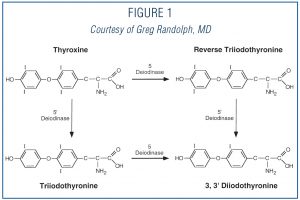
The thyroid’s job is to make thyroid hormones, which are secreted into the blood and then carried to every tissue in the body. Thyroid hormone helps the body use energy, stay warm and keep the brain, heart, muscles, and other organs working as they should (Figure 1).

THYROID-SURGERY-1

GENERAL INFORMATION

Thyroid operations are advised for patients who have a variety of thyroid conditions, including both cancerous and benign (non-cancerous) thyroid nodules, large thyroid
glands (goiters), and overactive thyroid glands. There are several thyroid operations that a surgeon may perform, including:

  1. excisional biopsy – removing a small part of the thyroid gland (rarely in use today);
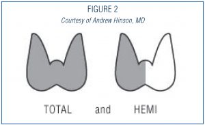
  2. lobectomy – removing half of the thyroid gland (the most frequent way to remove a nodule); (Figure 2)
  3.  total thyroidectomy, which removes all identifiable thyroid tissue. (Figure 2)
    There are specific indications for each of these operations.
    The main risks of a thyroid operation involve possible damage to important structures near the thyroid, primarily the parathyroid glands (which regulate calcium levels) and the recurrent and external laryngeal nerves (which control the vocal cords).


THYROID-SURGERY-2

QUESTIONS AND CONSIDERATIONS

When thyroid surgery is recommended, patients should ask several questions regarding the surgery including:

  1. Why do I need an operation?
  2. Are there other means of treatment?
  3. How should I be evaluated prior to the operation?
  4. How do I select a surgeon?
  5. What are the risks of the operation?
  6. How much of my thyroid gland needs to be removed?
  7. Will I need to take a thyroid pill after my operation?
  8. What can I expect once I decide to proceed with surgery?
  9. What will be my physical restrictions following surgery?
  10. Will I lead a normal life after surgery?

WHY DO I NEED AN OPERATION?

The most common reason for thyroid surgery is to remove a thyroid nodule, which has been found to be suspicious through a fine needle aspiration biopsy (see Thyroid Nodule brochure). Surgery may be recommended for the following biopsy results:

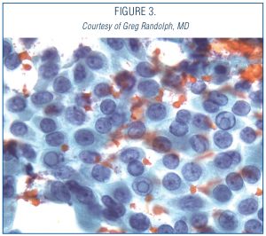
  1. cancer (papillary cancer); (Figure 3)
  2.  possible cancer (follicular neoplasm or atypical findings); or
  3.  inconclusive biopsy;
  4. molecular marker testing of biopsy specimen which indicates a risk for malignancy.


Thyroid Surgery

Surgery may be also recommended for nodules with benign biopsy results if the nodule is large, if it continues to increase in size or if it is causing symptoms (pain, difficulty swallowing, etc.). Surgery is also an option for the treatment of hyperthyroidism (Grave’s disease or a “toxic nodule”, for large and multinodular goiters and for any goiter that may be causing symptoms.

THYROID-SURGERY-3

ARE THERE OTHER MEANS OF TREATMENT?

Surgery is definitely indicated to remove nodules suspicious for thyroid cancer. In the absence of a possibility of thyroid cancer, there may be nonsurgical options of therapy depending on the diagnosis. You should discuss other options for therapy with your physician who has expertise in thyroid diseases.

HOW SHOULD I BE EVALUATED PRIOR TO THE OPERATION?

As for other operations, all patients considering thyroid surgery should be evaluated preoperatively with a thorough and comprehensive medical history and physical exam including cardiopulmonary (heart) evaluation. An electrocardiogram and a chest x-ray prior to surgery are often recommended for patients who are over 45 years of age or who are symptomatic from cardiac disease. Blood tests may be performed to determine if a bleeding disorder is present.
Any patients who have had a change in voice or who have had a previous neck operation (thyroid surgery, parathyroid surgery, spine surgery, carotid artery surgery, etc.) and/or who have suspected invasive thyroid disease should have their vocal cord function evaluated preoperatively. This is necessary to determine whether the recurrent laryngeal nerve that controls the vocal cord muscles is functioning normally and is becoming a norm of practice. Finally, if medullary thyroid cancer is suspected, patients should be evaluated for coexisting adrenal tumors (pheochromocytomas) and for hypercalcemia and hyperparathyroidism.

HOW DO I SELECT A SURGEON?

In general, thyroid surgery is best performed by a surgeon who has received special training and who performs thyroid surgery on a regular basis. The complication rate of thyroid operations is lower when the operation is done by a surgeon who does a considerable number of thyroid operations each year. Patients should ask their referring physician where he or she would go to have a thyroid operation or where he or she would send a family member.

WHAT ARE THE RISKS OF THE OPERATION?

The most serious possible risks of thyroid surgery include:

  1.  Bleeding that can cause acute respiratory distress,
  2.  Injury to the recurrent laryngeal nerve that can cause permanent hoarseness, and breathing problems with possible tracheotomy in rare cases if injury is sustained on both sides and
  3.  Damage to the parathyroid glands that control calcium levels in the body, causing hypoparathyroidism and hypocalcemia.

These complications occur more frequently in patients with invasive tumors or extensive lymph node involvement, in patients requiring a second thyroid surgery, and in patients with large goiters that go below the collarbone. Overall the risk of any serious complication should be less than 2%. However, the risk of complications discussed with the patient should be the particular surgeon’s risks rather than that quoted in the literature. Prior to surgery, patients should understand the reasons for the operation, the alternative methods of treatment, and the potential risks and benefits of the operation (informed consent).

HOW MUCH OF MY THYROID GLAND NEEDS TO BE REMOVED?

Your surgeon should explain the planned thyroid operation, such as lobectomy (hemi) or total thyroidectomy, and the reasons why such a procedure is recommended. For patients with papillary or follicular thyroid cancer many, but not all, surgeons recommend total or near total thyroidectomy when they believe that subsequent treatment with radioactive iodine might be beneficial. For patients with large (>1.5 cm) or more aggressive cancers and for patients with medullary thyroid cancer, more extensive lymph node dissection is necessary to remove possibly involved lymph node metastases.

Thyroid lobectomy may be recommended for overactive one-sided nodules or for benign one-sided nodules that are causing symptoms such as compression, hoarseness, shortness of breath or difficulty swallowing. A total or near – total thyroidectomy may be recommended for patients with Graves’ Disease or for patients with enlarged multinodular goiters

WILL I NEED TO TAKE A THYROID PILL AFTER MY
OPERATION?

The answer to this depends on how much of the thyroid gland is removed. If half (hemi) thyroidectomy is performed, there is an 80% chance you will not require a thyroid pill UNLESS you are already on thyroid medication for low thyroid (Hashimoto’s thyroiditis). If you have your entire (total) or remaining (completion) thyroidectomy, then you have no internal source of thyroid hormone remaining and you will need lifelong thyroid hormone replacement.

WHAT CAN I EXPECT ONCE I DECIDE TO PROCEED
WITH SURGERY?

Once you have met with the surgeon and decided to proceed with surgery, you will be scheduled for your pre-op evaluation (see above) and will meet with the anesthesiologist (the person who will put you to sleep during the surgery). You should have nothing to eat or drink after midnight on the day before surgery and should leave valuables and jewelry at home. The surgery usually takes 2-2½ hours, after which time you will slowly wake up in the recovery room. Surgery may be performed through a standard incision in the neck or may be done through a smaller incision with the aid of a video camera (Minimally invasive video assisted thyroidectomy).

There may be a surgical drain in the incision in your neck (which will be removed after the surgery) and your throat may be sore because of the breathing tube placed during the operation. Once you are fully awake, you will be moved to a bed in a hospital room where you will be able to eat and drink as you wish. Many patients having thyroid operations are hospitalized for about 24 hours and can be discharged on the morning following the operation.

WHAT WILL BE MY PHYSICAL RESTRICTIONS FOLLOWING SURGERY?

Most surgeons prefer a brief limitation is extreme physical activities following surgery. This is primarily to reduce the risk of a post operative neck hematoma (blood clot) and breaking of stitches in the wound closure. These limitations are brief, usually followed by a quick transition back to unrestricted activity. Normal activity can begin on the first postoperative day. Vigorous sports, such as swimming, and activities that include heavy lifting should be delayed for at least ten days to 2 weeks.

WILL I BE ABLE TO LEAD A NORMAL LIFE AFTER SURGERY?

Yes. Once you have recovered from the effects of thyroid surgery, you will usually be able to doing anything that you could do prior to surgery. Some patients become hypothyroid following thyroid surgery, requiring treatment with thyroid hormone. This is especially true if you had your whole thyroid gland removed. Thyroid hormone replacement therapy might be delayed for several weeks if you are to receive radioactive iodine (RAI) therapy unless there is a plan for you to receive TSH injection prior to RAI.

Read More

THYROID NODULES

WHAT IS THE THYROID GLAND?

The thyroid gland is a butterfly-shaped endocrine gland that is normally located in the lower front of the neck. The thyroid’s job is to make thyroid hormones, which are secreted into the blood and then carried to every tissue in the body. Thyroid hormone helps the body use energy, stay warm and keep the brain, heart, muscles, and other organs working as they should.

WHAT IS A THYROID NODULE?

The term thyroid nodule refers to an abnormal growth of thyroid cells that forms a lump within the thyroid gland. Although the vast majority of thyroid nodules are benign (noncancerous), a small proportion of thyroid nodules do contain thyroid cancer. In order to diagnose and treat thyroid cancer at the earliest stage, most thyroid nodules need some type of evaluation.

WHAT ARE THE SYMPTOMS OF A THYROID
NODULE?

Most thyroid nodules do not cause symptoms. Often, thyroid nodules are discovered incidentally during a routine physical examination or on imaging tests like CT scans or neck ultrasound done for completely unrelated reasons. Occasionally, patients themselves find thyroid nodules by noticing a lump in their neck while looking in a mirror, buttoning their collar, or fastening a necklace. Abnormal thyroid function tests may occasionally be the reason a thyroid nodule is found. Thyroid nodules may produce excess amounts of thyroid hormone causing hyperthyroidism (see Hyperthyroidism brochure).

However, most thyroid nodules, including those that cancerous, are actually non-functioning, meaning tests like TSH are normal. Rarely, patients with thyroid nodules may complain of pain in the neck, jaw, or ear. If a nodule is large enough to compress the windpipe or esophagus, it may cause difficulty with breathing, swallowing, or cause a “tickle in the throat”. Even less commonly, hoarseness can be caused if the nodule invades the nerve that controls the vocal cords but this is usually related to thyroid cancer.

The important points to remember are the following:

  • Thyroid nodules generally do not cause symptoms.
  • Thyroid tests are most typically normal—even when cancer is present in a nodule.
  • The best way to find a thyroid nodule is to make sure your doctor checks your neck!

WHAT CAUSES THYROID NODULES AND HOW COMMON ARE THEY?

We do not know what causes most thyroid nodules but they are extremely common. By age 60, about one-half of all people have a thyroid nodule that can be found either through examination or with imaging. Fortunately, over 90% of such nodules are benign. Hashimoto’s thyroiditis, which is the most common cause of hypothyroidism, is associated with an increased risk of thyroid nodules.

HOW IS A THYROID NODULE EVALUATED AND DIAGNOSED?

Once the nodule is discovered, your doctor will try to determine whether the rest of your thyroid is healthy or whether the entire thyroid gland has been affected by a more general condition such as hyperthyroidism or hypothyroidism. Your physician will feel the thyroid to see whether the entire gland is enlarged and whether a single or multiple nodules are present. The initial laboratory tests may include measurement of thyroid hormone (thyroxine, or T4) and thyroid-stimulating hormone (TSH) in your blood to determine whether your thyroid is functioning normally. Since it’s usually not possible to determine whether a thyroid nodule is cancerous by physical examination and blood tests alone, the evaluation of the thyroid nodules often includes specialized tests such as thyroid ultrasonography and fine needle biopsy.

THYROID ULTRASOUND:

Thyroid ultrasound is a key tool for thyroid nodule evaluation. It uses high-frequency sound waves to obtain a picture of the thyroid. This very accurate test can easily determine if a nodule is solid or fluid filled (cystic), and it can determine the precise size of the nodule. Ultrasound can help identify suspicious nodules since some ultrasound characteristics of thyroid nodules are more frequent in thyroid cancer than in noncancerous nodules. Thyroid ultrasound can identify nodules that are too small to feel during a physical examination. Ultrasound can also be used to accurately guide a needle directly into a nodule when your doctor thinks a fine needle biopsy is needed. Once the initial evaluation is completed, thyroid ultrasound can be used to keep an eye on thyroid nodules that do not require surgery to determine if they are growing or shrinking over time. The ultrasound is a painless test which many doctors may be able to perform in their own office.

THYROID FINE NEEDLE ASPIRATION BIOPSY (FNA OR FNAB):

A fine needle biopsy of a thyroid nodule may sound frightening, but the needle used is very small and a local anesthetic may not even be necessary. This simple procedure is often done in the doctor’s office. Sometimes, medications like blood thinners may need to be stopped for a few days before to the procedure. Otherwise, the biopsy does not usually require any other special preparation (no fasting). Patients typically return home or to work after the biopsy without even needing a bandaid! For a fine needle biopsy, your doctor will use a very thin needle to withdraw cells from the thyroid nodule. Ordinarily, several samples will be taken from different parts of the nodule to give your doctor the best chance of finding cancerous cells if they are present. The cells are then examined under a microscope by a pathologist.

The report of a thyroid fine needle biopsy will usually indicate one of the following findings:

  1. The nodule is benign (noncancerous).
    • This result is obtained in up to 80% of biopsies. The risk of overlooking a cancer when the biopsy is benign is generally less than 3 in 100 tests or 3%. This is even lower when the biopsy is reviewed by an experienced pathologist at a major medical center. Generally, benign thyroid nodules do not need to be removed unless they are causing symptoms like choking or difficulty swallowing. Follow up ultrasound exams are important. Occasionally, another biopsy may be required in the future, especially if the nodule grows over time.
  2. The nodule is malignant (cancerous) or suspicious for malignancy .
    • A malignant result is obtained in about 5% of biopsies and is most often due to papillary cancer, which is the most common type of thyroid cancer. A suspicious biopsy has a 50-75% risk of cancer in the nodule. These diagnoses require surgical removal of the thyroid after consultation with you docrinologist and surgeon.
  3. The nodule is indeterminate. This is actually a group of several diagnoses that may occur in up to 20% of cases. An Indeterminate finding means that even though an adequate number of cells was removed during the fine needle biopsy, examination with a microscope cannot reliably classify the result as benign or cancer.
    • The biopsy may be indeterminate because the nodule is described as a Follicular Lesion. These nodules are cancerous 20-30% of the time. However, the diagnosis can only be made by surgery. Since the odds that the nodule is not a cancer are much better here (70-80%), only the side of the thyroid with the nodule is usually removed. If a cancer is found, the remaining thyroid gland usually must be removed as well. If the surgery confirms that no cancer is present, no additional surgery to “complete” the thyroidectomy is necessary.
    • The biopsy may also be indeterminate because the cells from the nodule have features that cannot be placed in one of the other diagnostic categories.This diagnosis is called atypia, or a follicular lesion of undetermined significance. Diagnoses in this category will contain cancer rarely, so repeat evaluation with FNA or surgical biopsy to remove half of the thyroid containing the nodule is usually recommended.
  4. The biopsy may also be nondiagnostic or inadequate. This result is obtained in less than 5% of cases when an ultrasound is used to guide the FNA.
    • This result indicates that not enough cells were obtained to make a diagnosis but is a common result if the nodule is a cyst. These nodules may require reevaluation with second fine needle biopsy, or may need to be removed surgically depending on the clinical judgment of your doctor.

NUCLEAR THYROID SCANS:

Nuclear scanning of the thyroid was frequently done in the past to evaluate thyroid nodules. However, use of thyroid ultrasound and biopsy have proven so accurate and sensitive, nuclear scanning is no longer considered a first-line method of evaluation. Nuclear scanning still has an important role in the evaluation of rare nodules that cause hyperthyroidism. In this situation, the nuclear thyroid scan may suggest that no further evaluation or biopsy is needed. In most other situations, neck ultrasound and biopsy remain the best and most accurate way to evaluate all types of thyroid nodules.

MOLECULAR DIAGNOSTICS:

Can any other tests assist in evaluation of thyroid nodules? Yes, new tests that examine the genes in the DNA of thyroid nodules are currently available and more are being developed. These tests can provide helpful information about whether cancer may be present or absent. These tests are particularly helpful when the specimen evaluated by the pathologist is indeterminate. These specialized tests are done on samples obtained during the normal biopsy process. There are also specialized blood tests that can assist in the evaluation of thyroid nodules. These are currently available only at highly specialized medical centers, however, their availability is increasing rapidly. Ask your doctor if these tests are available and might be helpful for evaluating your thyroid nodule.

HOW ARE THYROID NODULES TREATED?

All thyroid nodules that are found to contain a thyroid cancer, or that are highly suspicious of containing a cancer, should be removed surgically by an experienced thyroid surgeon. Most thyroid cancers are curable and rarely cause life-threatening problems. Thyroid nodules that are benign by FNA or too small to biopsy should still be watched closely with ultrasound examination every 6 to 12 months and annual physical examination by your doctor. Surgery may still be recommended even for a nodule that is benign by FNA if it continues to grow, or develops worrisome features on ultrasound over the course of follow up.

Read More

MASTECTOMY OR LUMPECTOMY

MASTECTOMY OR LUMPECTOMY — FOR BREAST CANCER; OR BREAST BIOPSY FOR LUMPS OR ABNORMAL MAMMOGRAMS

What Is Breast Cancer?

Breast cancer is the abnormal growth and uncontrolled division of cells in the breast. Cancer cells can invade and destroy surrounding normal tissue, and can spread throughout the body via blood or lymph fluid (clear fluid bathing body cells) to start growing in another part of the body. Breast cancer is the most common malignancy among women. There are several types of breast cancer, distinguished mostly by their rate of growth and tendency to spread to other organs. Breast cancer often spreads to surrounding lymph nodes under the armpit, under the sternum (breastbone), and under the clavicle (collarbone).

Who gets it and what are its causes?

Every woman is at risk for breast cancer. As a woman ages, her risk of developing breast cancer rises dramatically regardless of her family history. The breast cancer risk of a 25 year-old woman is only one out of 19,608; by age 45, it is one in 93. In fact, 80% of all breast cancers are found in women over age 50.

Breast cancer also affects men. Symptoms, diagnosis, treatments and living with the disease apply to men as well as to women. Experts do not recommend routine screenings for men. Men account for 1% of cases, where in contrast, women face a one in ten lifetime risk of developing breast cancer.

There are a number of risk factors for the development of breast cancer. Family history of breast cancer in a mother or sister, early onset of menstruation and late menopause, reproductive history in regard to women who had no children or have children late in life, women who have never breastfed and those with a history of abnormal breast biopsies. However, more than 70% of women who get breast cancer have no known risk factors. In addition, some studies suggest that high fat diets, bottle feeding instead of breastfeeding or using alcohol may contribute to the risk profile.

Not all lumps detected in the breast are cancerous. Many are benign and require only the removal of the lump. While having several risk factors may boost a woman’s chances of having breast cancer, the relationship of factors is complex. The precise causes of breast cancer are unknown. The best way to assess breast cancer risk is by having clinical exams or doing regular self-examinations to detect any lump at an early stage. The second is to have a regular mammogram.

What are the symptoms?

Early detection and treatment are extremely important in curing breast cancer. The warning signs and symptoms of breast cancer could include the following:

Change in the size or shape of the breast

Lump or thickening of tissue in the breast or armpit

Dimpled or pulling of the skin over the breast

Nipple discharge

Retraction of the nipple

Scaliness of the nipple

Pain or Tenderness

Abnormality on a mammogram

Diagnosis

When breast cancer is found and treated early, the chances for survival are better. Women can take an active part in the early detection of breast cancer by having regular screening mammograms, clinical breast exams (breast exams performed by health professionals) and by performing breast self-exams.

Treatment

Mammogram

More than 90% of all breast cancers are detected by a mammogram (a low-dose x-ray of the breast). Mammography remains the best way of detecting signs of breast cancer. A baseline mammogram should be done by age 40, so that a normal x-ray can be used to compare future mammograms, even when there is no reason to believe there is a lump or cyst. Mammograms for women between the ages of 40 and 50 should be at the direction of her physician, usually every 1-2 years. An annual mammogram is recommended for women over the age of 50, and for most women with have suspicious breast lumps.

Radiation

This treatment is used after the cancer has been removed. The doctor may recommend radiation to destroy or shrink any remaining breast cancer cells. Radiation stops the cancer cells from dividing. It works especially well on fast-growing tumors. Unfortunately, it also stops some types of healthy cells from dividing. Healthy cells that divide quickly, like those of the skin, are affected the most. This is why radiation can cause skin problems.

Chemotherapy

Breast cancer surgery may be followed by chemotherapy, even in the earliest stages. Chemotherapy is administered either by injection of a blood vessel or orally. It is usually given in cycles, followed by a period of times for recovery and then followed by another course of drugs. Treatment time may range between four to nine months.

There may be significant side effects with some types of chemotherapy, including nausea and vomiting, temporary hair loss, mouth sores, vaginal sores, fatigue, weakened immune system and infertility. However, chemotherapy for early breast cancer uses medications that cause few side effects.

Hormone therapy

The growth of some breast cancer cells may be slowed by using the drug tamoxifen. If given each day as a pill, tamoxifen travels throughout the bloodstream, affecting all cells in the body. Tamoxifen treatment lasts between two to five years. Research suggests that tamoxifen may lower the chance that a breast cancer can return by between 25% and 35%.

Side effects of tamoxifen may include a slightly higher risk of endometrial cancer (cancer in the lining of the uterus). The risk increases if the drug is taken for more than five years. Other side effects include menopause-like symptoms like weight gain, hot flashes and mood swings.

Other possible hormone treatments include the use of estrogens, androgens and progestins. In rare cases, surgeons may suggest removal of the ovaries in premenopausal women as a way of eliminating the main source of estrogen, which can boost the growth of some breast tumors.

Surgical Treatment

Breast biopsy

A breast biopsy is the removal of breast tissue for examination by a pathologist. This can be accomplished surgically, or by withdrawing tissue through a needle.

A needle biopsy removes part of the suspicious area for examination. There are two types, aspiration biopsy (using a fine needle), and large core needle biopsy. Either of these may be called a percutaneous (procedure done through the skin) needle biopsy.

The fine needle biopsy uses a very thin needle to withdraw fluid and cells that can be studied. This can be done in your doctor’s office. No specialized equipment is needed. However, the use of an ultrasound helps to insure the guidance of the needle and that the specimen is taken from the right place.


A large core needle biopsy uses a larger diameter needle to remove small pieces of tissue, about the size of a grain of rice. It can be done in the rooms, a clinic or hospital. Ultrasound or x-ray is used for guidance of a large core needle biopsy.

A biopsy is recommended when a significant abnormality is found, either on physical examination and/or by an imaging test. Examples of abnormality can include a breast lump felt during physical examination and/or by an imaging test. It may also include a breast lump felt during a physical self-examination or tissue changes noticed from a mammogram test.

Before a biopsy is performed, it is important to make sure that the threat of cancer cannot be disproved or ruled-out by a simpler, less invasive examination. It may be obvious that a lump is harmless when examined by an ultrasound.

Mastectomy

The surgical removal of the breast for the treatment or prevention of breast cancer.

There are four types of mastectomies: partial mastectomy, or lumpectomy, in which the tumor and surrounding tissue is removed; simple mastectomy, where the entire breast is removed; modified radical mastectomy, in which the entire breast and all axillary lymph nodes are removed; and the radical mastectomy, where the entire breast, axillary lymph nodes, and chest muscles are removed.

The size, location, and type of tumor are very important when choosing the best surgery to treat a woman’s breast cancer. The size of the breast is also an important factor. A woman’s psychological concerns, and her lifestyle choices should also be considered when decisions are made.

The severity of a cancer is evaluated according to a complex system called Staging. This takes into account the size of the tumor, and whether it has spread to the lymph nodes, adjacent tissues and/or distant parts of the body. A mastectomy is usually the recommended surgery for more advanced breast cancers. Women with earlier stage breast cancers, could have breast-conserving surgery (lumpectomy).

There are many factors that make a mastectomy the treatment of choice for a patient. A large tumor is often an indication of a later stage of breast cancer, when the removal of the entire breast is recommended. In addition, large tumors are difficult to remove with good cosmetic results. This is especially true if the woman has small breasts. Sometimes multiple areas of cancer are found in one breast(called Multicentricity), making removal of the whole breast necessary. A cancer that has already attached itself to nearby tissues, such as the skin or chest wall, is most likely to be removed with a mastectomy. Immediate reconstruction post-mastectomy should always be considered.


Breast conserving surgery (Lumpectomy) are done for earlier stage breast cancers. Under certain conditions this may be insufficient,- eg multicentricity, incomplete resection, positive margins –  leading to completion Mastectomy. The surgeon is sometimes unable to remove the tumor with a sufficient amount or margin of normal tissue surrounding it. The entire breast needs to be removed in this situation. Some larger tumors can be treated with chemotherapy first (neo-adjuvant chemotherapy) to shrink the tumor, making Lumpectomy possible instead of Mastectomy.

Radiation therapy is always recommended following a lumpectomy. If a woman is unable to have radiation, a mastectomy is the treatment of choice. Pregnant women cannot have radiation therapy, for fear of harming the fetus. A woman with certain collagen vascular diseases, such as systemic lupus erythematosus or scleroderma, would experience unacceptable scarring and damage to her connective tissue from radiation exposure. Any woman who has had therapeutic radiation to the chest area for other reasons cannot tolerate additional exposure for breast cancer therapy. Diminished lung capacity due to other diseases also makes a woman a poor candidate for radiation therapy.

Prophylactic mastectomy, or removal of the breast to prevent future breast cancer, is controversial. Women with a strong family history of breast cancer and/or who test positive for a known cancer-causing gene may choose this option. Patients who have had certain types of breast cancers that are more likely to recur may elect to have the unaffected breast removed. Although there is some evidence that this procedure can decrease the chances of developing breast cancer, it is not a guarantee. It is not possible to be certain that all breast tissue has been removed. There have been cases where breast cancers have occurred after both breasts have been removed.

The decision to have mastectomy or lumpectomy should be carefully considered.

It is important that the woman be fully informed of all the potential risks and benefits of different surgical treatments before making a choice.

Read More

BREAST CANCER

MASTECTOMY OR LUMPECTOMY — FOR BREAST CANCER; OR BREAST BIOPSY FOR LUMPS OR ABNORMAL MAMMOGRAMS

What Is Breast Cancer?

Breast cancer is the abnormal growth and uncontrolled division of cells in the breast. Cancer cells can invade and destroy surrounding normal tissue, and can spread throughout the body via blood or lymph fluid (clear fluid bathing body cells) to start growing in another part of the body. Breast cancer is the most common malignancy among women. There are several types of breast cancer, distinguished mostly by their rate of growth and tendency to spread to other organs. Breast cancer often spreads to surrounding lymph nodes under the armpit, under the sternum (breastbone), and under the clavicle (collarbone).

Who gets it and what are its causes?

Every woman is at risk for breast cancer. As a woman ages, her risk of developing breast cancer rises dramatically regardless of her family history. The breast cancer risk of a 25 year-old woman is only one out of 19,608; by age 45, it is one in 93. In fact, 80% of all breast cancers are found in women over age 50.

Breast cancer also affects men. Symptoms, diagnosis, treatments and living with the disease apply to men as well as to women. Experts do not recommend routine screenings for men. Men account for 1% of cases, where in contrast, women face a one in ten lifetime risk of developing breast cancer.

There are a number of risk factors for the development of breast cancer. Family history of breast cancer in a mother or sister, early onset of menstruation and late menopause, reproductive history in regard to women who had no children or have children late in life, women who have never breastfed and those with a history of abnormal breast biopsies. However, more than 70% of women who get breast cancer have no known risk factors. In addition, some studies suggest that high fat diets, bottle feeding instead of breastfeeding or using alcohol may contribute to the risk profile.

Not all lumps detected in the breast are cancerous. Many are benign and require only the removal of the lump. While having several risk factors may boost a woman’s chances of having breast cancer, the relationship of factors is complex. The precise causes of breast cancer are unknown. The best way to assess breast cancer risk is by having clinical exams or doing regular self-examinations to detect any lump at an early stage. The second is to have a regular mammogram.


What are the symptoms?

Early detection and treatment are extremely important in curing breast cancer. The warning signs and symptoms of breast cancer could include the following:

Change in the size or shape of the breast

Lump or thickening of tissue in the breast or armpit

Dimpled or pulling of the skin over the breast

Nipple discharge

Retraction of the nipple

Scaliness of the nipple

Pain or Tenderness

Abnormality on a mammogram

Diagnosis

When breast cancer is found and treated early, the chances for survival are better. Women can take an active part in the early detection of breast cancer by having regular screening mammograms, clinical breast exams (breast exams performed by health professionals) and by performing breast self-exams.

Mammogram

More than 90% of all breast cancers are detected by a mammogram (a low-dose x-ray of the breast). Mammography remains the best way of detecting signs of breast cancer. A baseline mammogram should be done by age 40, so that a normal x-ray can be used to compare future mammograms, even when there is no reason to believe there is a lump or cyst. Mammograms for women between the ages of 40 and 50 should be at the direction of her physician, usually every 1-2 years. An annual mammogram is recommended for women over the age of 50, and for most women with have suspicious breast lumps.

Breast biopsy

A breast biopsy is the removal of breast tissue for examination by a pathologist. This can be accomplished surgically, or by withdrawing tissue through a needle.

A needle biopsy removes part of the suspicious area for examination. There are two types, aspiration biopsy (using a fine needle), and large core needle biopsy.

The fine needle aspiration uses a very thin needle to withdraw fluid and cells that can be studied. This can be done in your doctor’s office. No specialized equipment is needed. However, the use of an ultrasound helps to insure the guidance of the needle and that the specimen is taken from the right place.

A large core needle biopsy uses a larger diameter needle to remove small pieces of tissue, about the size of a grain of rice. It can be done in the rooms, a clinic or hospital. Ultrasound or x-ray is used for guidance of a large core needle biopsy.

A biopsy is recommended when a significant abnormality is found, either on physical examination and/or by an imaging test. Examples of abnormality can include a breast lump felt during physical examination and/or by an imaging test. It may also include a breast lump felt during a physical self-examination or tissue changes noticed from a mammogram test.

Before a biopsy is performed, it is important to make sure that the threat of cancer cannot be disproved or ruled-out by a simpler, less invasive examination. It may be obvious that a lump is harmless when examined by an ultrasound.

Surgical Treatment

Mastectomy

The surgical removal of the breast for the treatment or prevention of breast cancer.

There are four types of mastectomies: partial mastectomy, or lumpectomy, in which the tumour and surrounding tissue is removed; simple mastectomy, where the entire breast is removed; modified radical mastectomy, in which the entire breast and all axillary lymph nodes are removed; and the radical mastectomy, where the entire breast, axillary lymph nodes, and chest muscles are removed.

The size, location, and type of tumour are very important when choosing the best surgery to treat a woman’s breast cancer. The size of the breast is also an important factor. A woman’s psychological concerns, and her lifestyle choices should also be considered when decisions are made.

The severity of a cancer is evaluated according to a complex system called Staging. This takes into account the size of the tumour, and whether it has spread to the lymph nodes, adjacent tissues and/or distant parts of the body. A mastectomy is usually the recommended surgery for more advanced breast cancers. Women with earlier stage breast cancers, could have breast-conserving surgery (lumpectomy).

There are many factors that make a mastectomy the treatment of choice for a patient. A large tumour is often an indication of a later stage of breast cancer, when the removal of the entire breast is recommended. In addition, large tumours are difficult to remove with good cosmetic results. This is especially true if the woman has small breasts. Sometimes multiple areas of cancer are found in one breast(called Multicentricity), making removal of the whole breast necessary. A cancer that has already attached itself to nearby tissues, such as the skin or chest wall, is most likely to be removed with a mastectomy. Immediate reconstruction post-mastectomy should always be considered.

What is breast reconstruction?

In some cases it is possible to reconstruct the breast that has been removed, although the end result will not feel like your original breast. Reconstruction will not be able to give back the exact appearance and shape of your original breast. There are several different kinds of reconstruction. These have different recovery times and will cause different levels of discomfort. Sometimes reconstruction can be done at the same time as the mastectomy operation (immediate reconstruction) but sometimes a second operation is offered at a later stage (delayed reconstruction). With some reconstructions further surgery is needed to match the appearance of the reconstructed breast with the original breast. This may include operating on the unaffected breast to achieve symmetry. Your surgeon will talk with you about these options and give you some more information if reconstruction is suited to your case and is something that you would like to consider.

Breast conserving surgery (Lumpectomy) are done for earlier stage breast cancers. Under certain conditions this may be insufficient,- e.g. multicentricity, incomplete resection, positive margins –  leading to completion Mastectomy. The surgeon is sometimes unable to remove the tumour with a sufficient amount or margin of normal tissue surrounding it. The entire breast needs to be removed in this situation. Some larger tumours can be treated with chemotherapy first (neo-adjuvant chemotherapy) to shrink the tumour, making Lumpectomy possible instead of Mastectomy.

In most cases, an axillary node sample (to remove some lymph nodes in the armpit) or an axillary node clearance (to remove all of the lymph nodes in the armpit) may be performed at the same time as the lumpectomy. These operations are done to assess whether the cancer has spread to any of the lymph nodes (also called glands) as this information helps to plan any further treatments you may need. If these operations are suggested then your surgeon will explain which is best for you and why. Alternately you may be offered sentinel lymph node biopsy to check that the lymph nodes are clear – again if this is recommended for you, then you will be given some more information about it.

Radiation therapy is always recommended following a lumpectomy. If a woman is unable to have radiation, a mastectomy is the treatment of choice. Pregnant women cannot have radiation therapy, for fear of harming the foetus. A woman with certain collagen vascular diseases, such as systemic lupus erythematosis or scleroderma, would experience unacceptable scarring and damage to her connective tissue from radiation exposure. Any woman who has had therapeutic radiation to the chest area for other reasons cannot tolerate additional exposure for breast cancer therapy. Diminished lung capacity due to other diseases also makes a woman a poor candidate for radiation therapy.

Prophylactic mastectomy, or removal of the breast to prevent future breast cancer, is controversial. Women with a strong family history of breast cancer and/or who test positive for a known cancer-causing gene may choose this option. Patients who have had certain types of breast cancers that are more likely to recur may elect to have the unaffected breast removed. Although there is some evidence that this procedure can decrease the chances of developing breast cancer, it is not a guarantee. It is not possible to be certain that all breast tissue has been removed. There have been cases where breast cancers have occurred after both breasts have been removed.

The decision to have mastectomy or lumpectomy should be carefully considered.

It is important that the woman be fully informed of all the potential risks and benefits of different surgical treatments before making a choice.

What are the risks of this operation?

Possible risks and complications include:

  • Bleeding from the stitches or inside the wound. You should not be concerned if you find a small amount of blood spotting your wound dressing, but if more bleeding than this occurs after your discharge you should contact your surgeon
  • Infection. If your wound becomes inflamed, red, hot, sore or oozes pus you should contact your surgeon.
  • Thrombosis. This is a risk with all surgery and occurs when a blood clot forms in a vein, usually in the leg. You may be given blood thinning (anticoagulation) injections and you will be advised to wear support stockings whilst in hospital to help prevent this.
  • Numb areas in the arm/shoulder or pins and needles. Surgery can cause damage to the nerves – some of this will improve over time, although the scar itself will remain numb permanently.
  • Immobility or a ‘frozen’ shoulder. This is caused by not moving the arm following surgery. You will be given information on gentle exercises and advice on preventing this.
  • Risk of lymphoedema. This is swelling of the arm caused by the surgery or radiotherapy. You will be given information on exercises and advice on preventing this.

How will I recover from the operation?

The operation itself takes about one and a half hours and is under a general anaesthetic. After the operation time is spent in the Recovery Room until you are awake enough to return to the ward. You may find that you have an intravenous infusion or ‘drip’ in your arm for a few hours. This is to give you fluids directly into a vein until you feel able to drink, usually later on in the same day.

Expect to feel sore for a few days. Regular painkillers will be offered on a regular basis. If these are not effective please inform the nursing staff so that alternative pain relief can be offered to you.

What are drains?

Drains are plastic tubes, which allow blood and fluid to drain away from the wound and collect in a bottle or bag. Not everyone will have a drain inserted. Others may have one or two drains coming from under the wound. Drains also help to minimise bruising. If drains are used, the length of time until they are removed will vary from 7-10 days – your surgeon will be happy to discuss this with you in more detail.

When can I return home?

This depends on whether or not you have drains, or want to go home with a drainage bag or bottle still in place. If you would prefer to go home with your drains in place, it is often possible to go home on the day following the operation. If you decide that you do not want to go home with the drain, you may be in hospital for three to five days.

How should I care for the wound?

Your wound will be covered with a waterproof dressing and you will be able to bath or shower as usual during this time. You may find bathing relieves discomfort and helps you move your arm. Most surgeons use dissolvable stitches (sutures) which do not require removal, but if stitches or clips need removing, this will be done around 10 days after the operation. Steri-strips (little strips of plaster) may be used to give extra support to the wound. You can get these wet and they will start to loosen after about 10 days when they can be eased off as you would a plaster. Many people find it difficult to look at the wound, especially in the early days. However, being able to look at the wound seems to be a way of helping in the adjustment and acceptance process. You may prefer to have someone with you when you first look at the operation site.

What will happen after discharge?

Although adjustment may not be easy after the operation, be kind to yourself and take time to recover. The length of time needed to rest and recover after this operation depends very much on you as an individual. There are no real restrictions on what you may or may not do, but heavy lifting is not advised for at least six weeks. You can expect to feel a little sore for a few days. You will be offered pain killers regularly. If these are not effective, please tell the nurses or doctors. If your lymph nodes have been removed you may have a numb feeling on the inside of your arm. Sometimes it can be quite painful. It does improve with time, although some areas sometimes remain numb.

Any drainage tubes that have been used will be removed usually 7-10 days after your operation. This can be a little uncomfortable and some patients find it painful. Some women experience a ‘seroma’ which is a collection of fluid underneath the arm or under the wound. It may be uncomfortable but is not harmful. If a seroma develops, and is causing you concern, you may wish to visit your surgeon. He can advise you whether you will need to have the fluid drained.

When can I drive?

You can drive as soon as you can make an emergency stop without discomfort in the wound. This may be about 10 days after the operation. You must also be comfortable wearing a seatbelt.

You should speak to your insurance company about any restrictions following surgery.

What exercises should I do?

Arm exercises should be performed regularly after the operation to encourage the full range of movement back to your arm and shoulder. We suggest you perform the exercises three or four times each day after taking some pain relieving medication to allow easier movement.

You should continue with the exercises given to you in hospital until you feel that your arm and shoulder movements are back to normal. Some women prefer to continue these exercises indefinitely to prevent any problems developing. There is no reason why gentle exercise (such as swimming) should not be resumed as soon as you feel comfortable, usually about three or four weeks after surgery. More strenuous exercise can be resumed when your own doctor advises.

Removal of the lymph nodes from your armpit can leave a small numb area right up in the armpit which can be permanent. Numbness on the inner part of the arm usually returns to normal within a few weeks and you may feel some pins and needles while it is healing. This feeling should not restrict your mobility in any way.

What follow up treatment will I have?

This will be discussed at your follow-up appointment when the results from your operation and your treatment plans are finalised. As part of your follow up care it is likely you will be seen by an oncologist. In some cases surgery is all that is necessary. In others, further treatment is advised to reduce the risk of recurrence. Further treatment can include:

Radiotherapy

Radiotherapy is often advised after breast surgery. This is the use of high energy X-rays to destroy any remaining cancer cells. . Radiation stops the cancer cells from dividing. It works especially well on fast-growing tumours. Unfortunately, it also stops some types of healthy cells from dividing. Healthy cells that divide quickly, like those of the skin, are affected the most. This is why radiation can cause skin problems. Radiotherapy is localised treatment to the chest wall. It is not usually painful and you will not be radioactive at any stage in the treatment. Radiotherapy is given in specialist centres, as an outpatient treatment from Monday to Friday for about three weeks. This will be explained in more detail to you when you have your operation results discussed.

Endocrine treatment

Some breast tumours need the female hormone oestrogen to grow. A test will be done to see if you would benefit from anti-oestrogen tablets (oestrogen receptor). Tamoxifen or an aromatase inhibitor such as anastrazole or letrozole are commonly prescribed. There are a number of other similar tablets available and the choice will be discussed with you.

The tablets are taken daily and are prescribed for about five years. The main side effects are occasional hot flushes and muscle aches. These may be worse when you first start taking them. Generally the tablets should be trouble free and effective. Not all women will require these anti-oestrogen tablets. Note that these should not be confused with hormone replacement therapy (HRT) which should not be taken after a diagnosis of breast cancer.

Chemotherapy

This is treatment with anti-cancer drugs; the aim is to target cancer cells whilst doing the least damage to your normal cells. It is a systemic treatment which means that the drugs are usually injected into the blood stream and act throughout the body. The drug treatment may be given by injection, usually into a vein in the arm, or it can be given as tablets by mouth. is usually given in cycles, followed by a period of time for recovery and then followed by another course of drugs. Treatment time may range between four to nine months. There may be significant side effects with some types of chemotherapy, including nausea and vomiting, temporary hair loss, mouth sores, vaginal sores, fatigue, weakened immune system and infertility. However, chemotherapy for early breast cancer uses medications that cause few side effects.

Herceptin® (Trastuzumab)

Herceptin is a drug which can be given to some patients following their initial treatment. It can reduce the chance of breast cancer returning or slow down the rate of growth in an existing tumour. However it is only appropriate for around 1 in 4 patients who have a particular type of protein on their cancer cells. This is known as being ‘HER-2 positive’. When your breast cancer is diagnosed it will be tested to see if it is HER-2 positive and if this treatment is possible for you then you will be given some more information. If any of the treatments above are recommended for you, a more detailed explanation will be given. It may be that you are offered a combination of all of these treatments – this is common practice. It is important to remember that your medical team plan things differently for each individual, so try not to compare yourself to others.

Read More

BREAST BIOPSY

GENERAL INFORMATION

Thousands of women have breast masses that may cause them concern. These masses can be smaller than a pea or as large as an orange. There may be one mass or many masses. They can be solid or filled with fluid. Some are benign and others are likely to be cancerous; a lot of them are in between. Some masses can be felt whereas some are seen only on mammography. Because not all masses are the same, they do not all need to have a sample taken of them in the same way.

DIAGNOSIS

The majority of these breast lumps are not cancerous. If a risk of cancer is small your doctor may recommend careful observation. Generally, if the risk of cancer is thought to be greater than 2%, removing some of the suspicious tissue for testing is recommended. A biopsy can be done in one of several ways i.e. fine needle aspiration, needle core biopsy or open biopsy. After careful consideration of all the factors your doctor has recommended that you have a biopsy of the tissue in your breast and that this be done through a standard open incision.

PRE-OPERATIVE CARE

Your doctor will explain to you whether he will do the biopsy under local or general anaesthetic. If under general anaesthetic you must not have anything to eat or drink for 8 hours before the operation. Make sure your doctor knows what medicines you take, especially medicines which may affect blood clotting.

OPERATION

Under local or general anaesthetic your breast will be cleaned with an antiseptic. A cosmetically acceptable incision will be made. Tissue removed will be sent to the laboratory for examination. The bleeding in the wound will be stopped and the skin will be closed in an acceptable manner. Your doctor may or may not decide to leave a drain in the wound.

POST OPERATIVE CARE

You will initially be carefully observed in a recovery room and will be monitored. When you are stable you will be transferred back to your regular hospital bed. You may go home the same day.

HOME CARE

Your doctor will discuss post operative home activities with you. He will explain to you when you may resume normal activities, when you may drive and when you may return to work. Your doctor will discuss bathing and showering with you and will instruct you when to have the sutures removed.

COMPLICATIONS

  1. Complications are not common after a BREAST BIOPSY, but you must be aware of the following possible complications.
  2. A bleed or haematoma at the site of a BREAST BIOPSY does sometimes occur, even in spite of a drain having been left in. Depending on the extent of this bleed, this
    does sometimes need a repeat operation to evacuate the haematoma.
  3. The wound of a BREAST BIOPSY rarely becomes infected. If this happens, it may necessitate dressings or other active management.
  4. The scar after this operation is usually quite satisfactory, but some patients may end up with an unattractive scar.
  5. Systemic complications like venous thrombosis and pulmonary embolism are rare, but possible after any operation or anaesthetic.
Read More

HAEMORRHOIDECTOMY

GENERAL INFORMATION

Haemorrhoids are congested, swollen veins that are called internal haemorrhoids when they are just inside the anus. They are called external haemorrhoids when they are at the anal opening. There are many causes of haemorrhoids, including the following: chronic constipation, a job that requires a lot of sitting, pregnancy, liver disease, loss of muscle strength in the anal area with advancing age and obesity.

COMMON SIGNS AND SYMPTOMS

There can be a number of symptoms, sometimes more than one at the same time.

  • Protrusion of the haemorrhoids on the outside.
  • Pain or itching in and around the anus
  • Blood in the stool.
  • External haemorrhoids that are clotted and very painful.

DIAGNOSIS

• Usually the diagnosis can be made by taking a detailed history and doing a thorough physical examination.
• A painful, clotted haemorrhoid on the outside of the anus is easy to see.
• Anoscopy: This examination is done with a hollow instrument as long and as thick as your finger. It is lubricated and inserted into the anus gently so that the entire area can be inspected.

PRE-OPERATIVE PREPARATION

  • Do not eat or drink anything for 6 hours before the operation.
  • Your doctor will explain to you whether he wants you to have any laxatives or enemas before admission to hospital. He may prefer to have these given to you after you have been admitted.
  • Make sure your doctor knows what medicines you take, especially medicines which may affect blood clotting

OPERATION

  • The operation generally lasts between half an hour and one hour and will require a general anaesthetic.
  • A sigmoidoscopy or colonoscopy is generally done with a haemorrhoid operation. This is done to examine the lower part of the large intestine above the level of the anal canal. Your doctor will discuss this with you.

POST OPERATIVE CARE

  • Your doctor will discuss with you how long you need to stay in hospital.
  • At times the pain may be significant but your doctor will discuss with you how to manage this.
  • In addition to the pain there is always some post-operative bleeding. This is normal.

COMPLICATIONS

  1.  Complications are not common after a haemorrhoidectomy, but you must be aware of the following possible complications.
  2.  Occasionally a post operative bleed will occur. Your doctor will decide whether you need to be taken back to the operating theatre to manage this bleeding.
  3. A stricture or narrowing of the anus may develop occasionally after healing has taken place – this is extremely rare.
  4.  A temporary leak of flatus or faeces from the anus frequently persists for a few days after the operation. In very rare instances this problem may persist for a long time or even permanently.
  5. It is possible for further haemorrhoids to develop after a haemorrhoid operation.
  6. Systemic complications like deep vein thrombosis and pulmonary embolism are rare but possible after any operation or anaesthetic.
Read More

THORACOSCOPIC SYMPATHECTOMY

GENERAL INFORMATION
A sympathectomy is the removal of some of the sympathetic nerves which are responsible for control of the perspiration from the upper limb. This is done by means of an endoscope which is inserted through the chest cavity.

COMMON SIGNS AND SYMPTOMS
The disease hyperhidrosis results in excessive perspiration usually of the hands but sometimes also of the armpits

DIAGNOSIS
 The diagnosis is made clinically
 There are no tests to be done to make this diagnosis.
 Sometimes your doctor may decide to do a pre-operative chest X-ray

TREATMENT
 No creams or medication really cure or control this condition
 Botox injections are sometimes used but these give only temporary relief and can not cure this condition
 Your doctor has advised you to have a thoracoscopic sympathectomy.

PRE-OPERATIVE PREPARATION
 Make sure your doctor knows what medicines you take, especially medicines which may affect blood clotting.
 Do not eat or drink anything for 6 hours before the operation.
2/…
2.

OPERATION
 The operation will be done under general anaesthetic. Two or rarely three small incisions will be used on each side of your chest through which cannulae are inserted.
 An endoscope is used to view the sympathetic chain of nerves and the sympathetic ganglia at the back of the chest cavity by first collapsing the lung.
 This is a safer way of doing the operation than the previous way of doing the procedure through the neck.
 Two of the sympathetic ganglia are then divided or destroyed to interrupt the nerve impulses to the sweat glands of the hands. If the axilla is involved, then a third ganglion can be divided too.

POST OPERATIVE CARE
 After the operation you will be taken to a recovery room. When you are stable you will be taken to your regular hospital bed.
 Your doctor may decide to do a post-operative chest X-ray.
 Any pain, discomfort, or nausea will be controlled with medication.
 You can expect to be discharged a day or two after the operation.

COMPLICATIONS
 Serious complications are infrequent after a thoracoscopic sympathectomy. It is however important for you to remember that it is a major operation and complications may occur.
 Air may re-accumulate in your chest cavity after the operation forming a pheumothorax which may require a chest drain
 A bleed or an infection is always possible at the site of the small incisions.
 It is possible for the heart or lungs to be injured during this operation. This is rare. This may necessitate an open operation of the chest.
 A Horner’s syndrome with a drooping eyelid is possible but rare
 The operation may not be successful. The original hyperhidrosis can recur after months or years
 The biggest problem after this operation is that there is mostly a measure of compensatory hyperhidrosis. One does not really have control of this. It often manifests as increased perspiration from the body, the face or from the feet.
 Systemic complications like venous thrombosis and pulmonary embolism are rare, but possible after any operation or anaesthetic.

Read More

INCISIONAL HERNIA REPAIR

PATIENT INFORMATION ON INCISIONAL HERNIA

What is an Incisional Hernia?

An incisional hernia is a defect in your abdominal (belly) wall at the site of a previous incision (cut) which permits the intra-abdominal contents to push out. Incisional hernias occur when the old incision does not heal properly. It occurs more often in overweight patients, smokers, after emergency surgery, and when there was a wound infection. This can lead to your bowel being trapped which may cause an obstruction (blockage) or a strangulation (the blood supply to the bowel is cut off) of the contents. You may notice a swelling at the site of the incision (scar) of your previous operation – this can be associated with discomfort or pain in the abdomen, nausea and vomiting, and an inability to open bowels or pass wind.

Diagnosis of Incisional Hernia

An incisional hernia can cause you difficulty in daily activities like getting up from bed, dressing, walking, carrying shopping etc. It can cause symptoms like discomfort, pain, increasing size etc. Sometimes a loop of bowel can get trapped in the hernia and cause colicky abdominal pain or you may develop complications like bowel obstruction or strangulation. The diagnosis will usually be made by your doctor following a clinical examination if you have appropriate history and symptoms. Your GP will usually refer you to a surgeon – the surgeon may request investigations like an abdominal x-ray, a barium follow-through x-ray or a CT scan. Sometimes, you may not have time to see your GP because of the severity and acuteness of your symptoms, forcing you to attend the Accident and Emergency Department of your local hospital.

Management of Incisional Hernia

Incisional hernias come in different sizes. If you have a small incisional hernia and it is not causing any symptoms, then this can be initially kept under observation. However, if it is large or increasing in size or produces symptoms like discomfort, pain, nausea, vomiting or constipation etc. then a surgical repair is advisable, provided you are fit for surgery. If you live a sedentary life and the hernia is not causing any symptoms or increasing in size then then your doctor may advise continued observation only. The risk of this approach is that the hernia may increase in size in the future or develop an obstruction or strangulation which may require emergency surgery – the outcome under these circumstances may be worse than an elective operation. Please note that it is very likely that your hernia will enlarge over time, especially if you are physically active. Hence, it is advisable to have this repaired if you are generally fit to undergo surgery, especially if it is producing symptoms like discomfort, pain, nausea and constipation. The hernia repair is usually performed as a day case. You will be able to go home on the day of the operation. Occasionally, patients who expect to go home on the day of surgery may need to stay in overnight. Less often, for medical reasons or because of home circumstances, the operation will be done as an in-patient.

How is the Surgery Performed?

Open Incisional Hernia Repair

An incisional hernia can be repaired through the traditional open technique which requires a cut in the abdomen – Surgery involves return of the abdominal contents back into their normal position and repair of the area of weakness. A mesh (made from non-reactive material) is used to safely reinforce the area in repairing the overwhelming majority of hernias (both primary and recurrent hernias). The mesh used is a plastic net-like material. This sits in position forever supporting the weak area. The mesh does not react with normal tissues and causes no damage. You are likely to get aching and pulling during the first month after the operation. As you become more active, the tissues are stretched and become supple again. Mild twinges in the area of the hernia can continue for some months after the operation – no damage is being done.  The mesh patch becomes part of the body, giving strength and support to the abdominal wall. The mesh patch reduces, but does not eliminate, the chance of the hernia coming back.

Laparoscopic Incisional Hernia Repair

The hernia can is some cases also be repaired through a newer laparoscopic (keyhole surgery) technique which requires 4-5 small cuts in the abdomen (abdomen) – the trapped contents are released and the defect is closed with a mesh and staples from inside the abdominal cavity.

Risks of Incisional Hernia Surgery

All operations carry some risks which have to be weighed against the risks of complications if you do not undergo surgery. General risks of complications following an operation include risks of sore throat, pneumonia / atelectasis (collapsed lung), thrombo-embolism (clots in leg veins or in lungs), heart attack, stroke and death – these complications are rare.

The specific risks are different with the 2 techniques.

Open Incisional Hernia Repair

The wound is usually painful and there will be some swelling and discolouration which will usually settle with time. You may suffer from chronic wound pain, suffer an injury to structures, develop a leakage of bowel contents, intra-abdominal adhesions, a poor scar, seroma (body fluid collection), haematoma (blood clot collection), persistent obstruction, infection, recurrence of the hernia etc. – the risks of these happening are less than 5%.. You should be able to return to work within 4-6 weeks in most cases.

Laparoscopic Incisional Hernia Repair

The wounds are usually painful and there will be some swelling and discolouration which will usually settle with time. There is a 20% risk that your surgeon is unable to complete the operation laparoscopically and has to convert the procedure to open surgery due to intraoperative difficulty. You may suffer from chronic wound pain, suffer an injury to structures, develop a leakage of bowel contents, intra-abdominal adhesions, a poor scar, seroma (body fluid collection), haematoma (blood clot collection), persistent obstruction, infection, recurrence of the hernia etc. – the risks of these happening are less than 5%. You should be able to return to work within 2-6 weeks in most cases.

Advantages of key-hole operation

  • The recovery is quicker, so usually you can return to normal activity within 2-3 weeks.

Recovery is up to six weeks after an open repair

  • There is a much lower risk of long-term pain after a key-hole repair

Specific risks of the procedures

Infection         -A wound may become infected causing pain, redness and possibly discharge. The rate of risk is less than 2%.Infection is less common and less troublesome after a key-hole operation.

Bleeding         – Bleeding under the skin can produce a firm swelling – like a bruise. Some bleeding may occur in around 2% of operations.

The hernia recurs      – The hernia comes back. Further surgery for repair is required. The risk is up to 15%.

Swelling at the site of your previous hernia – There is often a swelling where the hernia had been, this is a localised bruise and not the hernia returned. This swelling settles over time

Seroma            – 5% – more common after laparoscopic repair

Unable to pass urine after operation

Damage to an internal structure – Rare (2%)

Some patients have discomfort from the staples for some months after keyhole surgery.

Pain is usually far less after a key-hole operation

Risks specific to the key-hole operation

Hernia at the site of instrument insertion

Need for open surgery – Keyhole surgery may not work and open surgery is needed – less

Outcome of Incisional Hernia Surgery

It is usual to experience some pain and discomfort for several weeks but this gradually subsides. Occasionally there may be a collection of clotted blood (haematoma) or body fluid (seroma) between the mesh and the skin – usually these are absorbed spontaneously though in a proportion of cases a further operation may be required. Following surgery there is resolution of symptoms in 95% of patients. You can sometimes experience significant pain after a repair – this is usually due to a nerve being trapped in the healing scar tissue. There is a 5% risk of you experiencing recurrent symptoms after some time following surgery requiring a further operation.

Returning to work:

You may return to light work as soon as you feel comfortable – this is usually possible within 2-4 weeks oafter the operation. You should avoid strenuous physical activity or heavy lifting for 4-6 weeks.

What happens if you decide not to undergo surgery for incisional hernia?

Some patients are content to continue observation until the hernia causes symptoms. However, if you do not undergo surgical repair of your hernia then you can experience progressive symptoms requiring an operation at a later date. The hernia may develop a blockage (intestinal obstruction) or develop strangulation (the blood supply to the bowel is cut off) which may require emergency surgery – the outcome under these circumstances may be worse than an elective operation. It is likely that a hernia will enlarge over time, especially in physically active persons.

WHAT HAPPENS AFTER THE OPERATION?

After surgery, you will be active and able to walk. Patients with small first-time hernia repairs are offered a light snack, and are usually discharged on the day of surgery safely and comfortably. Patients with larger, complex, or recurrent hernias will have to stay overnight in hospital. When you are discharged, a prescription for pain medication will be provided. Beware that pain medicines can cause drowsiness and constipation, so do not drive, eat lots of fruits and vegetables, drink plenty of water, and consider Metamucil or stool softeners while on pain medicine. You will have a waterproof dressing in place. Please leave it there until the first post-operative visit. I will remove it then. Discomfort, swelling, and bruising in the week or two after the operation are normal. Recovery and return to work and normal activity depends on how large or complex your hernia was and what type of work you do. You will be encouraged to be out of bed and walking within a few hours of the operation. This reduces the risk of complications like blood clots and pneumonia.

WHAT CAN I DO AFTER SURGERY?

You may shower the day after surgery. You can eat and drink whatever you like, unless I tell you otherwise. You can walk, climb stairs, and do light activity without delay. Activities such as jogging, tennis, and sexual activity can be resumed when your body feels comfortable doing them. You should not drive or operate heavy machinery as long as you need prescription pain medicine. I will discuss your expected recovery with you and give you specific instructions for return to heavier activity and work.

WHAT IF I DON’T HAVE THE HERNIA REPAIRED?

While hernias in babies sometimes heal, hernias in adults will never get better without surgery. In fact, they tend to enlarge and get worse over time. Many patients ask about a truss (external hernia support.) A truss may support the weak area and provide some comfort, but is generally ineffective and can cause pressure sores. Hernias can be aggravated by chronic cough, constipation, or heavy lifting. Contents of the hernia, like intestine, may occasionally become trapped within the hernia leading to intestinal blockage or damage ( incarceration or strangulation), creating an emergency surgical situation. If you notice constant severe pain at the site of the hernia, a lump that does not reduce in size when you lie down and relax, or symptoms such as pain with associated vomiting, you should call my office or report to a hospital emergency department without delay.

Read More